Auf unserer Website kommen verschiedene Cookies zum Einsatz: Technisch notwendige Cookies verwenden wir zu dem Zweck, Funktionen wie das Login oder einen Warenkorb zu ermöglichen. Optionale Cookies verwenden wir zu Marketing- und Optimierungszwecken, insbesondere um für Sie relevante und interessante Anzeigen bei den Plattformen von Meta (Facebook, Instagram) zu schalten. Optionale Cookies können Sie ablehnen. Mehr Informationen zur Datenerhebung und -verarbeitung finden Sie in unserer Datenschutzerklärung.
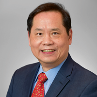
Hom-Lay Wang, DDS, MSD, PhD, is a professor and director of graduate periodontics at the University of Michigan. He has coedited two textbooks and published 50+ book chapters and 700+ scientific articles. He is co-editor-in-chief for Clinical Implant Dentistry and Related Research, associate editor for The International Journal of Oral & Maxillofacial Implants, a founding editorial board member for Clinical Advances in Periodontics, and vice president of the Academy of Osseointegration. Dr Wang is the recipient of numerous awards, including the American Academy of Periodontology Distinguished Service Award.
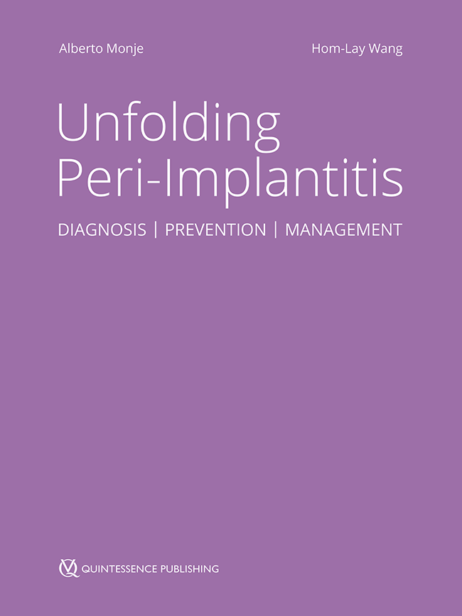
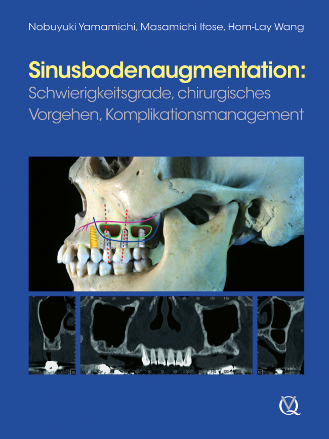
1. Auflage 2010 Buch Hardcover, 128 Seiten, 398 Abbildungen Sprache: Deutsch Kategorie: Implantologie Artikelnr.: 18010 ISBN 978-3-86867-012-7 QP Deutschland
Die gedruckte Ausgabe ist vergriffen. Bestellen Sie das E-Book über die Partner-Links.
eBooks
Veranstaltungen
The 14th International Symposium on Periodontics and Restorative Dentistry (ISPRD)
9. Juni 2022 — 12. Juni 2022Boston Marriott Copley Place, Boston, MA, Vereinigte Staaten von Amerika
Referenten: Tara Aghaloo, Edward P. Allen, Evanthia Anadioti, Wael Att, Vinay Bhide, Markus Blatz, Scotty Bolding, Lorenzo Breschi, Jeff Brucia, Daniel Buser, Luigi Canullo, Daniele Cardaropoli, Stephen J. Chu, Donald Clem, Christian Coachman, Lyndon F. Cooper, Daniel Cullum, Lee Culp, Sergio De Paoli, Marco Degidi, Nicholas Dello Russo, Serge Dibart, Joseph P. Fiorellini, Mauro Fradeani, Stuart J. Froum, David Garber, Maria L. Geisinger, William Giannobile, Luca Gobbato, Ueli Grunder, Galip Gürel, Chad Gwaltney, Christoph Hämmerle, Robert A. Horowitz, Markus B. Hürzeler, David Kim, Greggory Kinzer, Christopher Köttgen, Ina Köttgen, Purnima S. Kumar, Burton Langer, Lydia Legg, Pascal Magne, Kenneth A. Malament, Jay Malmquist, George Mandelaris, José Carlos Martins da Rosa, Pamela K. McClain, Michael K. McGuire, Mauro Merli, Konrad H. Meyenberg, Craig Martin Misch, Julie A. Mitchell, Marc L. Nevins, Myron Nevins, Michael G. Newman, Miguel A. Ortiz, Jacinthe M. Paquette, Stefano Parma-Benfenati, Michael A. Pikos, Giulio Rasperini, Pamela S. Ray, Christopher R. Richardson, Isabella Rocchietta, Marisa Roncati, Marco Ronda, Paul S. Rosen, Maria Emanuel Ryan, Irena Sailer, Maurice Salama, David M. Sarver, Takeshi Sasaki, Todd Scheyer, Massimo Simion, Michael Sonick, Sergio Spinato, Dennis P. Tarnow, Lorenzo Tavelli, Douglas A. Terry, Tiziano Testori, Carlo Tinti, Istvan Urban, Hom-Lay Wang, Robert Winter, Giovanni Zucchelli
Quintessence Publishing Co., Inc. USA
Zeitschriftenbeiträge dieses Autors
International Journal of Periodontics & Restorative Dentistry, Pre-Print
DOI: 10.11607/prd.6568, PubMed-ID: 37552187Seiten: 1-18, Sprache: EnglischWen, Shih-Cheng / Saleh, Muhammad / Alrmali, Abdusalam / Wu, David T / Wang, Hom-Lay
Despite the various barrier membranes proposed, one of the main challenges for guided bone regeneration (GBR) is space maintenance for large defects as well as ensure adequate blood supply. The presented feasibility case series aims to introduce an original titanium frame (TF) design, customized for each defect, as a modification of well-known principles and materials for GBR, for an enhanced and more predictable horizontal and vertical bone augmentation. Three patients with significant horizontal defects were treated with pre-trimmed TFs to create needed space, a 50%-50% mixture of autograft and bovine xenograft was placed, and then covered with collagen membrane. After 8 months of healing, the sites were reopened, the titanium screws were removed with the frame. An average of 8.0 ± 1.0mm horizontal and 3.0 ± 0.0mm vertical bone gain was achieved at the time of re-entry and implant placement surgery. Bone core biopsy was obtained during the implant placement. Histomorphometric analysis revealed that 42.8% of the sample was new vital bone, 18.8% was residual bone graft particles, and 38.4% was bone marrow like structures. After 3-4 months from implant placement, the implants were restored with provisional crowns and then finalized with zirconia screw-retained crowns. This case series suggests that GBR utilizing TFs with or without collagen membranes can be considered a suitable approach for horizontal and vertical bone augmentation. However, based on only three reported cases, the result should be carefully interpreted.
Successful bone augmentation relies on primary wound closure. Labial frenum is a soft tissue that connects the lip to alveolar mucosa or gingiva. However, frenum may exert biomechanical forces to the wound edge, causing wound instability. The aim of this study is to (1) review the frenum composition and classifications; (2) understand the significance of frenum in wound stability upon bone regeneration. An electronic search was conducted through the three online databases together with manual search on studies published until September 2022. A total of 300 articles were identified and 11 studies were included in this review. Two of the included six studies discovered that 35-37.5% of the labial frenum had muscle fibers. Other studies showed that labial frenum was mainly composed of connective tissue with elastic fibers. There are two widely used classifications for frenum based on morphology and position of attachment. No studies specifically evaluated the impact of frenum on bone regeneration. Frenum location intercorrelated with the amount of keratinized tissue, which could influence wound stability. A modified frenum classification for the edentulous ridge and a decision diagram to manage the frenum is proposed for research and evidenced practice.
Implants with deficient papillae and black triangle are common findings. The treatment of these esthetic complications is considered challenging, and with limited predictability. Therefore, the aim of the present report is to describe a novel technique for papilla augmentation (the "Iceberg" connective tissue graft [iCTG]) after extraction and interproximal bone reconstruction in the anterior region. A 35-year-old patient presented with a hopeless tooth with interproximal clinical attachment loss extending up the apical third of the adjacent tooth. Interproximal bone reconstruction was performed through alveolar ridge preservation by directly applying recombinant human platelet-derived growth factor-BB (rhPDGF-BB) to the exposed root surface of the adjacent tooth. A mixture of autogenous bone chips (obtained from the ramus) and bovine bone xenograft particles, previously mixed with the growth factor, was also used. The patient was able to come back for implant therapy only 2 years later. An incomplete regeneration of the interproximal bone was observed. Therefore, to compensate the interproximal deficiency, the iCTG approach, involving a double layer CTG with different origins, was utilized. Two small grafts from the tuberosity were sutured to the mesial and distal ends of a wider CTG harvested from the palate, aiming at gaining additional volume at the interproximal sites. The composite graft was then sutured on top of the implant platform, with the flap that was then released and closed by primary intention. After conditioning of the peri-implant tissues, the case was finalized with a satisfactory outcome. The described iCTG could be an effective approach for reconstructing peri-implant papilla following interproximal bone reconstruction.
Background: Multiple adjacent gingival recessions (MAGRs) are commonly treated with autogenous grafts. However, several intra- and post-surgical complications have been described following autogenous grafts, leading clinicians to explore the use of different biomaterials for the treatment of these conditions. The aim of the present study was to evaluate the root coverage outcomes of a novel porcine-derived acellular dermal matrix (PADM) in combination with the tunneled coronally advanced flap (TCAF) for the treatment of MAGRs.
Methods: Ten patients with 33 type 1 recession defects (RT1) were treated with PADM, in combination with the tunneled coronally advanced flap (TCAF). The outcomes of interest included the mean root coverage (mRC), the frequency of complete root coverage (CRC), changes in keratinized tissue width, volumetric gain at the treated sites assessed with digital intraoral scanning, as well as patient-reported outcome measures.
Results: All treated sites healed uneventfully, and no complications were noted throughout the study. At 6 months, a statistically significant reduction in recession was noted at the treated sites, exhibiting an overall mRC of 89.14 ± 19.15% and a CRC of 72.7%. The average volume gain after 6 months was 26.28 ± 11.71 in mm3 (Vol) and 0.63 ± 0.28 in mm (ΔD). The region-specific volumetric analysis revealed an overall higher linear dimensional gain at the Mid-Root aspect (ranging from 0.72 mm to 0.78 mm when assessed 1-4 mm apical to the cemento-enamel junction) compared to the other regions.
Conclusions: The present study presents the clinical and volumetric outcomes of PADM, in combination with TCAF for the treatment of MAGRs. A significant amount of volumetric gain was also observed as a result of the treatment at 6 months, along with satisfactory, esthetic and patient-reported outcomes.
Schlagwörter: Gingival recession, Acellular Dermal Matrix, Surgical flap, volumetric analysis, optical scanning
Extraction-site alveolar remodeling is a major concern due to negative volumetric hard and soft tissue changes that inevitably limit rehabilitation options and diminish esthetic outcomes. Surgical techniques employed to minimize alveolar dimensional changes are not always predictable. Utilizing socket-shield with immediate implant surgical procedure provides maintenance of a thin portion of the root in the vestibular area that minimizes bone resorption especially at the coronal cortical aspect. This case series assesses the dimensional changes in peri-alveolar structures via superimposition of the pre-operative and six-month post-operative 3D digital quantification of soft tissue. Fifty patients with 50 sites fulfilled the inclusion criteria. Implant survival was 100%, with no incidence of complications. Tissue changes were mesial papilla (-0.85mm), distal papilla (-0.95mm), vertical central margin (-0.7mm), horizontal central margin (-0.7mm), 4mm from the margin (-0.21mm), palatal central margin (-0.64mm). Clinically, the buccal contour was convex in 100% of the cases. The site with highest frequency to lose more than 1mm was the distal papilla, with 42% sites losing more than 1mm. The site with lowest frequency to lose more than 1mm was the point 4mm from the mid-facial margin, with 0% sites losing more than 1mm.
International Journal of Periodontics & Restorative Dentistry, Pre-Print
DOI: 10.11607/prd.6373, PubMed-ID: 37471156Seiten: 1-16, Sprache: EnglischChacón, Gerardo / Saleh, Muhammad H A / Decker, Ann / Kan, Joseph Y K / Wang, Hom-Lay
Successful rehabilitation of severely atrophic, short span edentulous ridges in esthetic can seldom be done without some form of vertical bone augmentation (VRA). The best available evidence shows guided bone regeneration (GBR) procedures may provide a very predictable option with a reduced potential for complications compared to alternative options. The present case series presents a novel technique to achieve predictable VRA with low complication tendency using tenting screws and cross-linked resorbable membranes. Ten patients with severe vertical defects in the esthetic zone participated in this study (5 males and 5 females; mean age of 35y). Following a mean healing time of 9.3 months, the mean defect resolution was 80%, with a mean vertical bone gain was 6.2mm (SD: 1.61 95% CI). Only one case presented with a reduced defect resolution (50%) of the defect, however, the bone gain for this case was 6mm.
Robotic systems have revolutionized various industries, and dentistry is no exception. Recently, due to the robust advancements in artificial intelligence and technology, there has been a significant evolution of dental robotic systems, ranging from surgeon controlled, robot-assisted operations, to more autonomous processes. The present clinical case report describes a 1-year follow-up of the successful use of an autonomous dental implant robot system with an osseodensification (OD) protocol for implant osteotomy preparation, maxillary sinus elevation, and simultaneous implant placement at the maxillary second premolar site. A prefabricated provisional prosthesis was delivered immediately after implant placement, with final prosthesis delivery at 3 months. The findings from this report demonstrate the integration and clinical augmentation of more autonomous protocols in the field of implant dentistry using dental robots.
Schlagwörter: Robotics surgery, Osseodensification, Maxillary sinus, Dental implant, Case report
This study clinically and histologically evaluated the outcome of a porcineapatite xenograft used to elevate the maxillary sinus floor in a severely atrophic ridge. A two-stage crestal window sinus elevation protocol was conducted in 24 patients with crestal bone ≤ 2 mm. Highly porous porcine carbonate apatite moistened with saline solution was placed in the elevated sinus cavity as the sole grafting material. Bone core biopsy samples were taken at 6, 9, and 12 months after sinus augmentation surgery (at implant placement). Treatment outcomes were assessed using microCT (μCT) and histologic analysis. Statistical analysis was performed using nonparametric Kruskal-Wallis test, followed by post-hoc Dunn multiple comparison test. At 6 months after implant placement, all implants achieved good primary stability (insertion torque ≥ 30 Ncm) and successfully osseointegrated. The residual graft amount (mean ± SE) was low (11.91% ± 1.99%) at 6 months and further decreased (6.11% ± 2.64%) by 12 months. On the contrary, the amount of new bone detected was 18.94% ± 4.08% at 6 months and was significantly (P < .05) increased (40.16% ± 5.27%) at 12 months. Histologic assessment revealed osteoclasts actively resorbing the graft as well as osteoblasts actively forming new bone. In the severely atrophic maxilla, the porcine-apatite xenograft promotes new bone formation while being slowly absorbed. Within the limited sample size, the porcine-apatite xenograft seems to be a good graft material for crestal window sinus augmentation.
This evaluation correlates maxillary sinus 3D morphology with bone regeneration. In 39 patients with crestal bone ≤ 2 mm, mineralized human bone allografts were used to augment the sinus floor through the crestal window sinus elevation approach. CBCT was used to measure the buccopalatal diameter (BPD), mesiodistal diameter (MDD), and hemi-ellipsoidal volume (Vh) in all sinuses. A bone core biopsy sample was taken at implant placement (4 to 5 months after sinus augmentation). Microradiographs of methacrylate-embedded sections were used to evaluate the amounts of bone, residual graft, and soft tissue. All 51 implants placed in the 39 patients successfully osseointegrated. A linear regression analysis showed that as BPD, MMD, and Vh increased, the amount of bone gain decreased and the amount of soft tissue increased (P < .05). The amount of residual graft was little affected by sinus morphology. Microradiographic data were grouped into four different sinus types (from small to great) using BPD and Vh medians. The best amount of bone formation was achieved in the narrow and short sinus type, while no great differences were found in the remaining three sinus types. Understanding of 3D sinus cavity morphology, especially the buccopalatal diameter and mesiodistal dimensions, is fundamental for achieving the best possible sinus augmentation outcomes.
Performing soft tissue augmentation (STA) at implant sites to improve esthetics, patient satisfaction, and peri-implant health is common. Several soft tissue grafting materials can be used to increase soft tissue thickness at the second-stage surgery, including human dermal matrices and xenogeneic collagen scaffolds. This study assessed and compared the volumetric outcomes, from second-stage surgery to crown delivery, around implants that received STA with a xenogeneic cross-linked collagen scaffold (XCCS) vs nonaugmented implant sites. Thirty-one patients (31 implant sites) completed the study. Intraoral digital scans were taken at the second stage and prior to crown delivery, and the STL files were imported in an image-analysis software to assess volumetric changes. XCCS-augmented implants showed significantly greater volumetric changes compared to control sites, which showed volume loss. The mean thickness of the XCCS-augmented area was 0.73 mm. There was no difference in patient-reported esthetic evaluations between groups. STA with XCCS provided significantly greater volumetric outcomes compared to nonaugmented sites. Further studies are needed to evaluate the long-term behavior of the augmented peri-implant mucosa and the effects of STA on peri-implant health.