Various cookies are used on our website: We use technically necessary cookies for the purpose of enabling functions such as login or a shopping cart. We use optional cookies for marketing and optimization purposes, in particular to place relevant and interesting ads for you on Meta's platforms (Facebook, Instagram). You can refuse optional cookies. More information on data collection and processing can be found in our privacy policy.
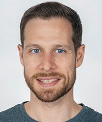
2006 bis 2011 Studium der Zahnmedizin an der Alber-Ludwigs-Universität Freiburg i. Br. 2012 bis 2016 Wissenschaftlicher Mitarbeiter Klinik für Zahnärztliche Prothetik des Departments für Zahn- Mund und Kieferheilkunde Freiburg. 2013 Promotion. 2016 Oberarzt an der Klinik für Rekonstruktive Zahnmedizin und Gerodontologie, Zahnmedizinische Kliniken, Universität Bern. 2018 Verantwortlicher für die postgraduierten Masterstudiengänge, Klinik für Rekonstruktive Zahnmedizin und Gerodontologie, Zahnmedizinsche Kliniken, Universität Bern. 2018 Leiter der Station für Digitale Implantat- und Rekonstruktive Zahnmedizin. Zusatzqualifikationen: Spezialist für Prothetik (DGPro), Fachzahnarzt für Rekonstruktive Zahnmedizin (Schweizerische Gesellschaft für Rekonstruktive Zahnmedizin; SSRD), Curriculum Implantologie (DGI) Forschungsschwerpunkte: Prothetische Versorgungskonzepte, Digitale Technologien, Gerodontologie
Events
The Buser & Belser Master Course on Esthetic Implant Dentistry
3 Live-Surgeries, 16 Lectures, 3 Hands-on Workshops (on-site only), 1 Social Dinner (on-site only)11. Oct 2023 — 13. Oct 2023SITEM Insel, Bern, Switzerland
Speakers: Samir Abou-Ayash, Urs C. Belser, Dieter Bosshardt, Daniel Buser, Jordi Caballé-Serrano, Stephen Chen, Vincent Fehmer, Manrique Fonseca, Ronald E. Jung, Irena Sailer, Anton Sculean
Buser & Sculean Academy
30th EAO Annual Scientific Meeting / 37th DGI Annual Congress
Speakers: Samir Abou-Ayash, Bilal Al-Nawas, Thomas Bernhart, Florian Beuer, Stefan Bienz, Elena Calciolari, Najla Chebib, Andreas Dengel, Vincent Donker, Joke Duyck, Roberto Farina, Gary Finelle, Alberto Fonzar, Tobias Fretwurst, Rudolf Fürhauser, Oscar Gonzalez-Martin, Stefano Gracis, Knut A. Grötz, Christian Hammächer, Lisa J. A. Heitz-Mayfield, Detlef Hildebrand, Norbert Jakse, Jim Janakievski, Tim Joda, Daniel Jönsson, Greggory Kinzer, Vincent G. Kokich, Michael Krimmel, Cecilia Larsson Wexell, Martin Lorenzoni, Georg Mailath-Pokorny, Julia Mailath-Pokorny, Frank Georg Mathers, Gerry McKenna, Henny Meijer, Alberto Monje, Torsten Mundt, Nadja Nänni, David Nisand, Robert Nölken, Nicole Passia, Michael Payer, Christof Pertl, Aušra Ramanauskaitė, Eik Schiegnitz, Martin Schimmel, Ulrike Schulze-Späte, Frank Schwarz, Falk Schwendicke, Robert Stigler, Michael Stimmelmayr, Anette Strunz, Christian Ulm, Stefan Vandeweghe, Kay Vietor, Arjan Vissink, Asaf Wilensky, Stefan Wolfart, Werner Zechner, Anja Zembic, Nicola Zitzmann
European Association for Osseintegration (EAO)
23. Prothetik Symposium
Machen, aber richtig!30. Nov 2019Pullmann Berlin Schweizerhof, Berlin, Germany
Speakers: Samir Abou-Ayash, Nadine Buchholz, Dominik Groß, Jan-Frederik Güth, Anita Hattenbach, Ursula Jasper, Ilka Johannemann, Friedhelm Klingenburg, Anja Liebermann, Simon Peroz, Jochen Peters, Sebastian Pflesser, Daniel Reinke, Ralph Riquier, Hans-Jürgen Stecher, Henry Theiling, Christian Vordermayer
Quintessenz Verlags-GmbH
This author's journal articles
International Journal of Periodontics & Restorative Dentistry, Pre-Print
DOI: 10.11607/prd.6626, PubMed ID (PMID): 37819849Language: EnglishFonseca, Manrique / Molinero-Mourelle, Pedro / Donmez, Mustafa Borga / Abou-Ayash, Samir / Buser, Daniel / Sculean, Anton / Yilmaz, Burak
Dental implants have been commonly used to replace missing single teeth. However, esthetic rehabilitation of an adjacent tooth may also be required due to diastemas, crowding, or existing large direct restorations to improve the final esthetic outcome. With the advancements in ceramics and bonding techniques, minimally invasive esthetic approaches have become viable for compromised spacing issues. This case report describes a dental technique for the esthetic rehabilitation of compromised anterior spacing with a customized zirconia implant abutment at maxillary central incisor site and a partial ceramic veneer bonded to adjacent central incisor.
Keywords: Anterior spacing; case report; implants; partial laminate veneer; prosthetic dentistry
Advanced digital technologies have rabidly been integrated into prosthodontics to improve the digital workflow for prosthetic rehabilitation. The integration of 3D datasets acquired from various imaging sources such as intraoral scanners and facial optical scanners allows the creation of virtual patients to perform presurgical simulation and prosthetic rehabilitation. The presented technique introduced a straightforward protocol for aligning intraoral scans (Trios 4, 3Shape) to optical face scans (Face Hunter, Zirkonzahn GmbH) using a global bestfit algorithm of 3D evaluation software (GOM Inspect). Nasal geometry data was used as a matching reference to produce virtual dental patients. This integration protocol proved that the intraoral scanner (Trios 4, 3Shape) was used not only to scan dental arches but also used effectively to scan the nose. These scans along with professional facial scans can be successfully aligned to produce virtual dental patients. Because only one single fully-dentate patient case with an alignment deviation of 243.6 μm was used, further research to evaluate the accuracy of this protocol is needed.
Keywords: Dental impression technique, face, imaging, nose, three-dimensional
Purpose: To evaluate 1-year survival and success rates of 6-mm short implants placed in mandibular molar sites with two different abutments (dome/ball) retaining existing removable partial dentures (RPDs).
Materials and Methods: In 19 patients, 38 implants of 6-mm length were placed bilaterally. After 4 months, each participant received the dome abutment, which 2 months later was exchanged with the ball abutment. Clinical data were recorded at abutment connection (4 months postsurgery) and at 6 and 12 months postsurgery, including probing depth, bleeding on probing, presence of plaque, and standardized radiographs. Implant success was assessed using the following criteria: presence of pain, mobility, radiographic bone loss, probing depth, and the presence of exudate. For descriptive analyses, mean and SD values were calculated. Paired sample t tests and linear regressions with a significance level of α < .05 were applied to analyze the evolution of peri-implant parameters and the influence of implant placement depth.
Results: The overall mean marginal bone level alteration (DMBL) was 1.05 ± 0.69 mm. A statistically significant marginal bone loss over time was observed at the mesial and distal aspects of all implants (P < .05). The implant survival rate was 100%. No implants showed pain, exudate, mobility, or probing depth > 7 mm. Three implants were classified as having satisfactory survival due to a DMBL > 2 mm (resulting success rate: 92.1%). No influence of implant placement depth was found.
Conclusion: These short-term results suggest that short implants can be used in mandibular molar sites for additional posterior support of free-end RPDs. However, in individual cases, DMBL > 2 mm may occur.
Purpose: To evaluate the effect of cooling on the reverse torque values of different abutments in bone-level and tissue-level implants. The null hypothesis was that there would be no difference in reverse torque values of abutment screws when cooled and uncooled implant abutments were compared.
Materials and Methods: Bone-level and tissue-level implants (Straumann, each n = 36) were placed in synthetic bone blocks and subdivided into three groups (each n = 12) based on the abutment type (titanium base, cementable abutment, abutment for screw-retained restorations). All abutment screws were tightened to 35 Ncm torque. In half of the implants, a dry ice rod was applied on the abutments close to the implant-abutment connection for 60 seconds before untightening the abutment screw. The remaining implant-abutment pairs were not cooled. The maximum reverse torque values were recorded using a digital torque meter. The tightening and untightening procedure was repeated three times for each implant including cooling for the test groups, resulting in 18 reverse torque values per group. Two-way analysis of variance (ANOVA) was used to analyze the effect of cooling and abutment type on the measurements. Post hoc t tests were used to make group comparisons (α = .05). The P values of post hoc tests were corrected for multiple testing using the Bonferroni-Holm method.
Results: The null hypothesis was rejected. Cooling and abutment type significantly affected the reverse torque values in bone-level implants (P = .004) but not in tissue-level implants (P = .051). The reverse torque values of bone-level implants significantly decreased after cooling (20.31 ± 2.55 Ncm vs 17.61 ± 2.49 Ncm). Overall mean reverse torque values were significantly higher in bonelevel implants compared to tissue-level implants (18.96 ± 2.84 Ncm vs 16.13 ± 3.17 Ncm; P < .001).
Conclusion: Cooling of the implant abutment led to a significant decrease in reverse torque values in bone-level implants and may therefore be recommended as a pretreatment before the application of procedures to remove a stuck implant part. Int J Oral Maxillofac Implants 2023;38:94–100. doi: 10.11607/jomi.9499
Keywords: abutment screw, blocked implants, cryo-mechanical, maintenance, technical complications
Purpose: To compare the load-to-failure values of different ceramic CAD/CAM implant crown materials with drilled screw-access holes with and without cyclic loading applied.
Materials and Methods: Forty zirconia abutments with a titanium base were pre-loaded onto implants to support maxillary right first premolar crowns milled from four different CAD/CAM ceramic materials (zirconia-reinforced lithium silicate, hybrid ceramic, lithium disilicate, and zirconia; n = 10 each). After cementing the crowns, the screw-access channels were prepared by drilling through the occlusal surfaces. Half of the specimens were subjected to cyclic loading for 5 million cycles at 2 Hz (n = 5/material). After cyclic loading, vertical loads were applied until failure. The load-to-failure values of all crowns were recorded and statistically analyzed. Two-way ANOVA was used with restricted maximum likelihood estimation and Tukey-Kramer adjustments (α = .05).
Results: During cyclic loading, the zirconia abutment under one lithium disilicate specimen cracked at 2 million cycles, and a zirconia-reinforced lithium silicate crown also fractured. Results for the load-to-failure test series showed statistical differences between the materials. Zirconia resulted in significantly higher failure loads when compared to the other materials (P < .001). Cyclic loading did not significantly affect the load-to-failure values.
Conclusion: Cyclic loading did not significantly influence the load to failure of any of the materials tested. Zirconia crowns with drilled screw access channels cemented on zirconia abutments with a titanium base had higher load-to-failure values compared to the other ceramic crown materials.
Purpose: To assess the effect of tooth morphology on the amount of tooth structure removal and the effect of different assessment methods on the detected amount of removed tooth structure.
Materials and Methods: Eight test groups (n = 10 each) of standardized artificial teeth were prepared for partial and full crowns. All teeth were prepared by the same operator following predefined preparation parameters. Tooth structure removal was measured by using three different assessment methods: digital volumetric analysis (DVA), weight analysis (WA), and combined computer-aided manufacture-weight analysis (CAMWA). Nonparametric repeated-measures ANOVA and post hoc analyses were used to determine the influence of tooth morphology and assessment method on the detected amount of tooth structure removal.
Results: For partial-crown preparations, only tooth morphology had a significant impact on the detected amount of tooth structure removal (P < .0001), but not the different assessment methods used (P = .08); tooth structure removal was not significantly different between the canine and incisor groups, but was significant for all other groupwise comparisons. For full-crown preparations, the tooth morphology (P = .047) and different assessment methods (P = .01) had an impact on the detected tooth structure removal; however, only a few groupwise comparisons reached the significance level.
Conclusion: The amount of tooth structure removal depended on tooth morphology and the type of assessment method, which should be taken into account when comparing results across studies. The detected amount of tooth structure removal was below the values described in the literature independent of the assessment method used.
Purpose: To investigate the volumetric facial soft tissue changes associated with wearing complete dentures using 3D facial scans.
Materials and Methods: A total of 40 volunteers (20 men and 20 women) were recruited for this study and were treated with maxillary and mandibular complete dentures. Six facial scans were taken of each subject; three scans wearing a complete denture and three without the complete denture. The 3D facial scans were taken with the mouth in three positions: closed, relaxed, and smiling. Each scan was superimposed in order to analyze and quantify linear measurements involving 14 soft tissue landmarks and the total volume that the subject gained with the prosthesis.
Results: Three variables were evaluated in each analysis: gender, mouth position, and age. In the analysis of the soft tissue landmarks, there was a significant effect of age, with patients > 75 years showing the greatest changes (P < .05). The landmarks that showed the most changes were those located around the mouth. In the volumetric analysis, the variable with the highest influence was gender, with men gaining more volume than women (P < .05).
Conclusion: Complete dentures have a significant effect on volumetric changes in perioral tissues. These changes are more marked in patients > 75 years. Compared to women, men depicted greater volumetric changes with complete dentures. These results provide a new avenue for clinicians and developers using facial scans to design future restorations for edentulous patients.
Der Fallbericht dokumentiert die konventionelle und die digitale Herstellungsmethode von Klammerprothesen. Es werden Chancen und Grenzen des digitalen Herstellungsverfahrens aufgezeigt. Zu den Chancen gehören der geringere Zeit- und Kostenaufwand, allerdings ist die Passgenauigkeit möglicherweise eingeschränkt.
Keywords: Klammerprothese, CAD/CAM, digitaler Workflow, Kommunikation, SLM
Aim: This case report describes a digital workflow for prosthetically driven surgical planning, implant placement, and the fabrication of two screw-retained, complete-arch, implant-supported dentures in an edentulous patient. The purpose of this case report is to present and discuss the digital steps in the workflow, especially the scanning technique to obtain the centric relation, through a clinical case. The limitations of the workflow are also discussed.Materials and methods: Static computer-aided implant surgery (s-CAIS) was planned three-dimensionally, based on cone beam computed tomography, intraoral optical scanning, and digital bite registration. Using s-CAIS, four and six implants were placed in the edentulous mandible and maxilla, respectively. The final screw-retained complete-arch monolithic zirconia restorations were manufactured based on a digital workflow, using the pre-existing modified radiologic guide for the digital maxillomandibular record. Conclusions: The evolution of digital processing methods allows for design, processing, and fabrication of implant-supported fixed complete dentures using a surgical, prosthetic, and technical workflow based on 3D restorative backward planning. A digital prosthetic setup and CAD/CAM can be used for the fabrication of intraoral try-ins that serve as a model for the final monolithic zirconia superstructure.
Keywords: backward planning, edentulous, complete-arch, implant-supported, fixed complete denture, CAD/CAM, monolithic zirconia, radiologic guide
Die Implantattherapie bei älteren Menschen bedarf einer gezielten Planung. Dabei spielt die Unterscheidung zwischen dem dritten und vierten Lebensabschnitt für die Behandlungsplanung und die anschließende zahnärztliche Betreuung eine wichtige Rolle, da diese Patientengruppen sich fundamental voneinander unterscheiden. Für Menschen des dritten Lebensabschnitts steht das gesamte Spektrum der zahnärztlichen Implantologie zur Verfügung, allerdings müssen chronische Erkrankungen, Medikation und manuelle Geschicklichkeit beachtet werden. Da dentale Implantate meist deutlich länger als 20 Jahre im Mund verbleiben, muss prospektiv die zukünftige Abnahme der biologischen, sozialen und kognitiven Funktionen bei der Planung berücksichtigt werden. Im vierten Lebensabschnitt ist das Leben von der Abhängigkeit bei den Aktivitäten des täglichen Lebens (ADL), Multimorbidität, Polypharmazie und Institutionalisierung bestimmt. Es gibt fast keine wissenschaftliche Evidenz zur Implantattherapie dieser Patientengruppe. Die wenigen vorhandenen Studien zeigen eine sehr hohe Implantatüberlebensrate und es gibt Hinweise auf verringerte akute periimplantäre Entzündungsreaktionen. Gerade Patienten in der vierten Lebensphase könnten am meisten vom funktionellen Gewinn einer implantatstabiliserten Totalprothese profitieren, wenn die muskulären Fähigkeiten zur Stabilisierung einer Prothese nachlassen. Allerdings müssen Handhabung, Nachsorge und tägliche Pflege gesichert werden, bevor mit einer solchen Therapie im Sinne des Nichtschadensgebots begonnen wird. Menschen im palliativen Kontext weisen zunehmend Implantate auf, die die Betroffenen und die Pflegenden vor große Herausforderungen stellen können. Hier müssen die Implantatprothesen rechtzeitig „rückgebaut“ werden, um Schmerzen und Infektionen zu vermeiden.
Manuskripteingang: 06.01.2021, Annahme: 19.02.2021
Keywords: Implantologie, Gerodontologie, dritter Lebensabschnitt, vierter Lebensabschnitt, chirurgische Strategien, prothetische Strategien