Wir verwenden Cookies ausschließlich zu dem Zweck, technisch notwendige Funktionen wie das Login oder einen Warenkorb zu ermöglichen, oder Ihre Bestätigung zu speichern. Mehr Informationen zur Datenerhebung und -verarbeitung finden Sie in unserer Datenschutzerklärung.
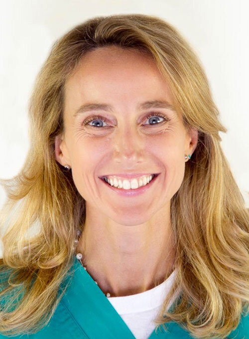
2005 schloss sie ihr Studium der Zahnmedizin an der Universität Bologna, Italien, ab. Sie promovierte 2016 an der Universität Bologna, Italien, in Medizinwissenschaften. Seit 2018 Forscherin an der Fakultät für biomedizinische und neuromotorische Wissenschaften der Universität Bologna. Mitglied des Redaktionsbeirats der Italienischen Gesellschaft für Parodontologie in 2018-2019. Mitglied des wissenschaftlichen Komitees der Italienischen Gesellschaft für Parodontologie in 2016/2017. Gastprofessur an der San Raffaele Universität Mailand 2015-2016. Dozentin im Postgraduierten-Studiengang der Universität San Raffaele Mailand 2017. Dozentin im zweiten Level des International Master an der Universität Bologna 2017/2018. Mitglied der NOGI (National Osteology Group Italy). Seit 2018 ITI Fellow, Study Club Director und Mitglied des ITI Leadership Development Committee. Seit 2012 aktives Mitglied der Italienischen Gesellschaft für Parodontologie (SIDP), von 2005 bis 2013 Forschungsstipendium bei Prof. Giovanni Zucchelli. Seit 2005 bis heute Zahnärztin in privater Praxis als Expertin für Parodontologie. Autorin mehrerer Publikationen in Pubmed. Co-Autorin eines Kapitels im Buch "Implant Therapy", herausgegeben von M. Nevins und H. L. Wang. Referentin auf nationalen und internationalen Kongressen zur Parodontologie.
1. Auflage 2022 Buch Hardcover; Two-volume book with slipcase; 21 x 28 cm, 1100 Seiten, 6557 Abbildungen Sprache: Englisch Kategorien: Oralchirurgie, Implantologie Artikelnr.: 7736 ISBN 978-88-7492-091-4 QP Italy
Erscheinungsweise: vierteljährlich Sprache: Deutsch Kategorie: Ästhetische Zahnheilkunde Schriftleitung / Chefredaktion: Dr. Martina Stefanini PhD, ZTM Vincent Fehmer BDT, MDT, Dr. Alfonso Gil DDS, PhD QP Deutschland
Erscheinungsweise: vierteljährlich Sprache: Englisch Kategorie: Ästhetische Zahnheilkunde Schriftleitung / Chefredaktion: Dr. Martina Stefanini PhD, ZTM Vincent Fehmer BDT, MDT, Dr. Alfonso Gil DDS, PhD QP Deutschland
Regeneration and Esthetics in Periodontology and Implant Dentistry02.12.2022 — 03.12.2022Congress Center, Kursaal Bern, Bern, Schweiz
Referenten: Sofia Aroca, Daniel Buser, Stephen Chen, Massimo De Sanctis, Nikolaos Donos, Markus B. Hürzeler, Sascha Jovanovic, Ronald E. Jung, Giulio Rasperini, Isabella Rocchietta, Frank Schwarz, Anton Sculean, Shakeel Shahdad, Andreas Stavropoulos, Martina Stefanini, Leonardo Trombelli, Istvan Urban, Giovanni Zucchelli
Buser & Sculean Academy
Zeitschriftenbeiträge dieses Autors
International Journal of Esthetic Dentistry (EN), 1/2023
PubMed-ID: 36734420Seiten: 9-10, Sprache: EnglischFehmer, Vincent / Gil, Alfonso / Stefanini, Martina
Autogenous soft tissue grafting is a commonly performed procedure in periodontal and implant surgery. Reharvesting a connective tissue graft (CTG) from the same palatal donor site is often required, but little is known about the volumetric changes that occur after harvesting a free gingival graft and how long the palatal mucosa takes to regain its original form and thickness. This study evaluated the volumetric changes that occur at the palatal donor site after harvesting a soft tissue graft with a noninvasive digital technology. Nineteen patients needing a CTG for a single site were enrolled. Intraoral digital scans of the palatal donor sites were obtained at baseline and at 1, 3, 6, and 12 months. The digital scans were imported and analyzed with an imaging software to evaluate volumetric changes. Average volume losses of 5.82 ± 2.63 mm3 and 11.03 ± 5.47 mm3 were observed after 1 and 3 months, respectively. Only minor changes were observed at 6 and 12 months. Linear dimensional changes at 5 and 7 mm from the gingival margin were substantially higher than the changes at 3 mm for the 1- and 3-month interval comparisons compared to baseline. Graft dimension was associated with volume loss at 1 and 3 months (P < .01). After palatal harvesting, the donor site undergoes volumetric changes, mostly during the first 3 months, and is attenuated thereafter.
Tooth malposition negatively affects the outcome of root coverage procedures, limiting chances for complete root coverage (CRC). This case series introduces a combined orthodontic-mucogingival approach for the treatment of deep (> 4 mm) isolated gingival recession defects affecting mandibular incisors with a buccally displaced root. Twenty patients were treated with a novel orthodontic device (FZ Root Torque Controller) for selective correction of tooth malposition at the affected site, turning all isolated recessions into Miller Classes I and II. Subsequent surgical treatment of the gingival recession with a vertically-coronally advanced flap plus a connective tissue graft achieved 90% CRC at the 1-year follow-up, with a recession reduction of 5.6 ± 1.5 mm, a 1.24 ± 0.24 mm increase in gingival thickness, and improved esthetic outcomes.
The gingival thickness (GT) and keratinized tissue width (KTW) constitute the gingival phenotype, a concept that has received a great deal of appreciation in recent years. Gingival phenotype modification has been achieved via different surgical techniques and grafting materials. Despite the superiority of autogenous grafts, their increased patient morbidity and limited recourse has led to the development of graft substitutes. The human dermal matrix is a notable example that, depending on its processing method, can become freeze-dried or solvent-dehydrated acellular dermal matrix (FDADM and SDADM, respectively). This article reports the 9-year outcomes of a randomized clinical trial regarding gingival phenotype modification following root coverage with FDADM and SDADM. Twelve of the original 20 patients were available at the 9-year follow-up. Overall, the outcomes of gingival phenotype modification were maintained in both groups and at all sites after 9 years. For KTW, an incremental increase was observed over time in both groups, and minimal or no changes were noted in GT from the 1-year recall to the 9-year recall. However, the gingival margin level showed an apical shift for both groups.
The coronally advanced flap technique is one of the most commonly used approaches for treating gingival recession. Several modifications of the technique have been proposed over the years, making it a highly predictable treatment option for gingival recession; however, as dental implants are structurally and biologically different from natural teeth, a further modification of the conventional coronally advanced flap technique has been suggested to overcome the challenges posed by the treatment of peri-implant soft tissue dehiscences. The present article aims to describe the state of the art of the technique at implant sites presenting with peri-implant soft tissue dehiscences, and highlight the main differences between this and the coronally advanced flap approach in natural teeth. The timing of crown removal and abutment modification/replacement are discussed, along with the different methods for management of the flap and connective tissue graft that are recommended at implant sites. The outcomes of this approach compared to the conventional coronally advanced flap technique, and other approaches are also presented.
Schlagwörter: connective tissue graft, dental implants, gingival recession, soft tissue augmentation, surgical flap
Conflict-of-interest statement: The authors do not have any financial interests, either directly or indirectly, in the products or information listed in the paper.
Treatment of gingival recessions affecting mandibular incisors is scarcely documented. Despite a shallow vestibule depth being considered a poor anatomical condition, it has never been measured nor deemed a clinical parameter affecting the outcome of root coverage procedures. This study describes a vertically and coronally advanced flap (V-CAF) + connective tissue graft (CTG) technique to obtain root coverage and increased vestibule depth in the treatment of gingival recessions affecting mandibular incisors. Twenty patients with single gingival recessions were treated. The results showed that V-CAF+CTG is effective in increasing residual vestibule depth and in reducing recession depth. Immediately after surgery, a vestibule-depth increase of 5.9 ± 1.2 mm was reported, which was statistically significant compared to baseline, and it remained stable after 12 months (4.8 ± 1.1 mm). The mean percentage of root coverage was 98.3% ± 5.2% for all treated recessions, and complete root coverage (CRC) was achieved in 90% of cases (18 of 20). V-CAF+CTG could be considered a successful technique in terms of vestibule depth increase and CRC for the treatment of single gingival recessions in the mandibular incisors.