International Journal of Computerized Dentistry, Pre-Print
ScienceDOI: 10.3290/j.ijcd.b4494409, PubMed-ID: 3782353912. Okt. 2023,Seiten: 1-31, Sprache: EnglischSchlenz, Maximiliane Amelie / Schulz-Weidner, Nelly / Olbrich, Max / Buchmann, Darlene / Wöstmann, Bernd
Aim: Even though today, many fields in dentistry allow digital processes, analogue procedures are still widely used. This cross-sectional pilot study aimed to survey insights on the digitalisation of dental practices using the example of Hesse.
Materials and Methods: Between April and June 2022, 4840 active practicing dentists registered by the State Dental Association of Hesse were invited via e-mail to fill out an online questionnaire regarding their technical requirements in dental practice, dental treatment procedures, and attitude towards digitalisation in dentistry. Demographic questions were asked. Besides descriptive statistics, correlations were analyzed (P < 0.05).
Result: Questionnaires of 937 dentists (279 female, 410 male, four inter/divers, 244 no answer; mean age of 51.4 ± 10.4 years) were examined representing a respond rate of 19.36%. In the area of practice administration and dental radiography, the majority of the dentists surveyed is already working digitally, which is predominantly assessed as a positive development. Already one third of the respondents state that they use an intraoral scanner for dental treatments, but the indication is mainly limited to smaller restorations. However, many dentists rate the use of social media accounts and telemedicine rather negative.
Conclusion: Within the limitation of this pilot study, many processes especially in dental treatments are still analogue. However, 60% of the participants plan digitalisation of their dental practices within the next five years, which indicates a clear shift from analogue to digital dentistry.
Schlagwörter: Analog-Digital Conversion, CAD/CAM, Dental Practice Pattern, Digital Technology, Intraoral Scanner, Organisation and Administration, Real World Data on Dentistry, Surveys and Questionnaires
The International Journal of Prosthodontics, Pre-Print
DOI: 10.11607/ijp.8843, PubMed-ID: 3853614822. März 2024,Seiten: 1-19, Sprache: EnglischSchmidt, Alexander / Berschin, Cara / Wöstmann, Bernd / Schlenz, Maximiliane Amelie
Purpose: To update data on the transfer accuracy of digital implant impressions by using a
coordinate-based analysis, latest intraoral scanners (IOSs) were investigated in an established
clinical close model set-up. Materials and Methods: An implant master model (IMM) of the
maxilla with four implants in the posterior area (#14/#24 and #16/#26) and a reference cube
was scanned with four different IOS (i700 (Medit), Primescan (Dentsply Sirona), Trios 4 and
Trios 5 (3Shape) ten times each. Datasets were compared with a reference dataset of IMM
that was generated with x-ray computed tomography in advance. 3D deviations for the
implant-abutment-interface points (IAIPs) were calculated. Statistical analysis was performed
by multifactorial ANOVA (p < .05). Results: Overall deviations for trueness (mean) ±
precision (SD) of the IAIPs ranged from 88±47 μm for the Primescan, followed by 112±57
μm for the i700, 121±42 μm for the Trios 4 and 124±43 μm for the Trios 5 with decreasing
accuracy along the scan path. For trueness, one significant difference between the Primescan
and the T4 was detected for one implant position. For precision, no significant differences
were noticed. Conclusions: Although the latest IOS showed a significant improvement in
transfer accuracy, the accumulating deviation along the scan path is not yet resolved.
Considering the Trios system, the innovation seems to be limited as no improvement could be
detected between Trios 4 and 5.
The International Journal of Oral & Maxillofacial Implants, Pre-Print
DOI: 10.11607/jomi.10612, PubMed-ID: 3838196713. Feb. 2024,Sprache: EnglischZierden, Karina / Reich, Sarah Marie / Vogler, Jonas Helmut Adrian / Wöstmann, Bernd / Rehmann, Peter
Purpose: This retrospective clinical follow-up study assesses double-crown retained implanttooth-
supported removable partial dentures (DCR-ITSRPDs) survival, evaluates abutment
survival and identifies first aftercare measures. Materials and Methods: The influence of various
factors on the survival of the DCR-ITSRPDs and the abutments were observed in this
retrospective clinical follow-up study using Kaplan-Meier estimate. In addition, the first occurred
aftercare measure per prosthesis was evaluated. Results: 47 DCR-ITSRPDs were investigated
(mean observation: 4.3 ± 3.8 years; max. 14.3 years) out of which three (6.4%) had to be
replaced. The 5- and 10- year survival probability for DCR-ITSRPDs was 100% and 75%. A total
of 297 abutments (120 natural teeth and 177 dental implants) were observed, of which 22 (7.4%;
6 teeth and 16 implants) failed. The 5- and 10-year survival probability for teeth was 90.2% and
for dental implants 90.4% and 76.3%. Conclusion: DCR-ITSRPDs are a successful and durable
treatment option for patients with substantially reduced residual dentitions. Both, prostheses and
abutments show good survival times after 5- and 10-years in function. The patient associated
factors tested showed no influence on the survival of DCR-ITSRPs and abutments. Peri implant
infection was the decisive factor for abutment loss, therefore, regular dental prophylaxis and
examinations are of major importance.
Schlagwörter: removable dental prosthesis, double crown, dental implants, tooth-implant supported, survival
The Journal of Adhesive Dentistry, 1/2023
Open Access Online OnlyResearchDOI: 10.3290/j.jad.b4051483, PubMed-ID: 37097056Seiten: 107-116, Sprache: EnglischHofmann, Maria / Amend, Stefanie / Lücker, Susanne / Frankenberger, Roland / Wöstmann, Bernd / Krämer, Norbert
Purpose: The aim of this in-vitro study was to evaluate the marginal integrity and wear of eight bulk-fill materials in comparison to a compomer in Class-II cavities in primary molars after thermomechanical loading (TML).
Materials and Methods: Prepared Class-II cavities in 72 extracted primary molars were filled with eight bulk-fill materials. A compomer served as the control group. After water storage (incubator, 28 days, 37°C), samples were subjected to TML (2500 thermal cycles 5°C/55°C; 100,000 load cycles, 50 N, 1.67 Hz). Before and after TML, replicas were made which were used for both SEM analysis of marginal integrity and 3-D wear analysis. Statistical analysis was performed using Kruskal-Wallis and Wilcoxon tests (p < 0.05).
Results: A significant reduction in perfect margins was observed for all groups, while marginal gap formation increased (Wilcoxon test, p < 0.02) for all groups but the compomer. Significant interindividual differences were observed between the tested materials regarding marginal integrity (Kruskal-Wallis test, p < 0.05). Wear analysis revealed no significant differences between groups (Kruskal-Wallis test, p > 0.05).
Conclusion: Some of the bulk-fill materials investigated here achieved better results than the compomer and should be further evaluated clinically.
Schlagwörter: Class-II restoration, composite, marginal analysis in SEM, polyacid modified resin, wear
QZ - Quintessenz Zahntechnik, 6/2022
KurzfassungSeiten: 640, Sprache: DeutschSchmidt, Alexander / Benedickt, Christopher R. / Schlenz, Maximiliane A. / Rehmann, Peter / Wöstmann, Bernd
The International Journal of Prosthodontics, 5/2022
DOI: 10.11607/ijp.7834Seiten: 690-696, Sprache: EnglischZierden, Karina / Wöstmann, Bernd / Rehmann, Peter
Purpose: To survey the clinical performance of telescopic-retained removable implant-supported dental prostheses (TR-RISDPs) in edentulous patients, as well as incidental maintenance measures and technical complications.
Materials and Methods: In this retrospective analysis, the former presence of oral cancer, prosthesis location (maxilla or mandible), and participation in a follow-up program were analyzed as possible factors with an influence on survival and maintenance treatments of the TR-RISDPs and dental implants using Kaplan-Meier estimates.
Results: A total of 86 TR-RISDPs (mean follow-up: 4.62 ± 3.24 years; maximum 13.8 years) and 465 implants (mean follow-up: 5.67 ± 3.59 years; maximum 16.5 years) were observed. Six (6.9%) of the TR-RISDPs had to be remade, and 11 (2.3%) implants failed. Regular attendance in the follow-up program showed significantly higher survival times and fewer maintenance treatments for the TRRISDPs (P < .05). Implants in patients with former oral cancer showed significantly lower survival times (P < .001).
Conclusions: TR-RISDPs in edentulous patients show excellent clinical outcomes. Regular check-ups are decisive for success.
The International Journal of Prosthodontics, 6/2021
DOI: 10.11607/ijp.6233Seiten: 756-762, Sprache: EnglischSchmidt, Alexander / Benedickt, Christopher R / Schlenz, Maximiliane A / Rehmann, Peter / Wöstmann, Bernd
Purpose: To evaluate the accuracy (trueness and precision) achievable with four intraoral scanners (IOSs) and different preparation geometries.
Materials and methods: A model of a maxillary arch with different preparation geometries (onlay, inlay, veneer, full-crown) served as the reference master model (RMM). The RMM was scanned 10 times using four commonly used IOSs (Trios 2 [TR], 3Shape; Omnicam [OC], Dentsply Sirona; True-Definition [TD], 3M ESPE; and Primescan [PS], Dentsply Sirona). Scans were matched using a 3D measurement software (Inspect 2019, GOM) and a best-fit algorithm, and the accuracy (trueness and precision) of the preparation types of the scanning data was evaluated for positive and negative deviations separately. All data were subjected to univariate analysis of variance using SPSS version 24 (IBM).
Results: Mean (± SD) positive deviations ranged from 4.6 ± 0.7 μm (TR, veneer) to 25.9 ± 2.4μm (OC, full crown). Mean negative deviations ranged from -7.2 ± 0.6 μm (TR, veneer) to -26.4 ± 3.8 μm (OC, full crown). There were significant differences (P < .05) in terms of trueness and precision among the different IOSs and preparation geometries.
Conclusion: The transfer accuracy of simple geometries was significantly more accurate than those of the more complex prosthetic geometries. Overall, however, the IOSs used in this study yielded results that were clinically useful for the investigated preparation types, and the mean positive and negative deviations were in clinically acceptable ranges.
The International Journal of Oral & Maxillofacial Implants, 5/2021
DOI: 10.11607/jomi.8784Seiten: 985-991, Sprache: EnglischZierden, Karina / Grau, Laura Sophie / Wöstmann, Bernd / Rehmann, Peter
Purpose: To investigate the clinical performance of implant-supported dental prostheses (ISDPs), this retrospective clinical study observed influencing factors on survival of the prostheses and necessary maintenance treatments during the observation time and complications of the dental implants.
Materials and methods: Patients who were provided either with fixed implant-supported dental prostheses (FISDPs) or telescopic-retained removable implant-supported dental prostheses (TR-RISDPs) were included in this retrospective clinical study. Potential influencing factors on the survival probability of the prostheses were observed using Kaplan-Meier analysis: patient sex, type of prosthesis, location, dentition in opposing arch, participation in follow-up visits, and whether the patient had a previous history of oral cancer. The type and number of maintenance treatments and complications of dental implants were also analyzed.
Results: A collective of 473 patients who were provided with either FISDPs (n = 320) or TR-RISDPs (n = 153) and 1,499 implants were included in the study. 6.6% of the prostheses (24 FISDPs and 7 TR-RISDPs) had to be replaced, and 6.3% of the implants (n = 45) were lost. The calculated 5-year survival probabilities were 87.4% for FISDPs and 95.5% for TR-RISDPs. FISDPs in patients who also had ISDPs in the opposing arch showed the lowest survival probabilities (P < .05). TR-RISDPs in patients who regularly attended follow-up visits showed the highest survival rates (P < .05). Maintenance treatments had to be performed at an earlier stage for patients with TR-RISDPs, and especially for TR-RISDPs located in the mandible (P < .05).
Conclusion: FISDPs and TR-RISDPs showed good survival rates in this study. However, when planning FISDPs, the dentition in the opposing arch should be considered to prevent possible failure. TR-RISDPs indicate a higher need for aftercare measures, especially in the early years of function. Regular attendance of follow-up visits is still a decisive factor for success.
Schlagwörter: dental implant, dental prosthesis, implant-supported, Kaplan-Meier estimate, retrospective studies
Implantologie, 3/2021
Seiten: 243-255, Sprache: DeutschWöstmann, Bernd / Schmidt, Alexander / Schlenz, Maximiliane Amelie
Insbesondere in der Implantologie eröffnet der intraorale Scan, neben der alleinigen Funktion einer Abformung, die Möglichkeit zur Implementierung neuer Behandlungskonzepte. Bereits in der Beratungs- und Planungsphase können so in Verbindung mit dreidimensionalen Röntgendaten Möglichkeiten, Grenzen und Risiken der Implantatversorgung erläutert und in einem prothetisch-chirurgischen Behandlungskonzept festgelegt werden, welches zu vorhersagbareren Behandlungsergebnissen führt. Jedoch müssen die heute noch bestehenden Limitationen der Ganzkieferversorgungen in Bezug auf die dreidimensionale Übertragung der Implantatposition von der Mundhöhle auf ein Modell beachtet werden, weshalb indikationsabhängig auch kombiniert digital-analoge Versorgungskonzepte in Betracht gezogen werden sollten. Durch die kontinuierliche Weiterentwicklung der Scansysteme ist jedoch zukünftig damit zu rechnen, dass auch hier die digitale Abformung die etablierten analogen Behandlungsverfahren ersetzen wird.
Manuskripteingang: 18.06.2021, Annahme: 11.08.2021
Schlagwörter: digitale Abformung, Implantatabformung, konventionelle Abformung, intraorale Scanner, Abformgenauigkeit, digitale Implantatplanung
International Journal of Computerized Dentistry, 2/2021
SciencePubMed-ID: 34085501Seiten: 157-164, Sprache: Englisch, DeutschSchmidt, Alexander / Billig, Jan-Wilhelm / Schlenz, Maximiliane Amelie / Wöstmann, Bernd
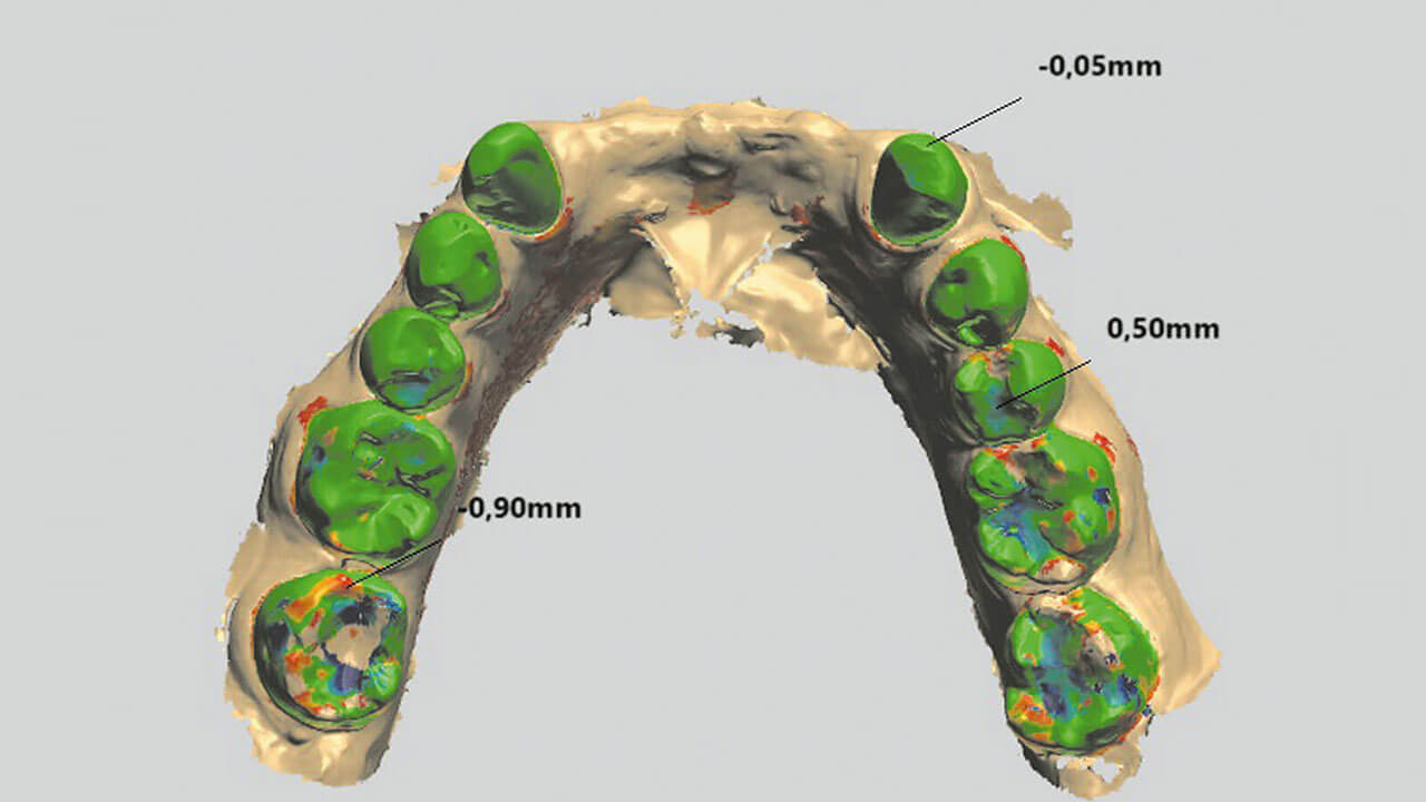
Ziel: Zur Bestimmung von metrischen Genauigkeiten, geht es innerhalb von zahnmedizinischen Genauigkeitsuntersuchungen typischerweise um die Bestimmung der Abweichungen zwischen Ist- und Soll-Datensätzen von Urmodellen. Dabei werden zur Analyse verschiedene Messmethoden verwendet, wobei die Ergebnisse häufig direkt miteinander verglichen werden. Ziel der vorliegenden Studie war es daher, den Einfluss und die Auswirkung verschiedener Methoden der digitalen Datenanalyse – koordinatenbasierte Analyse (CBA) und Best-Fit-Überlagerung – auf die Ergebnisse zu analysieren.
Material und Methode: Ein Modell mit vier Implantaten und einem Referenzquader wurde durch Mikro-Computertomografie (CT) digitalisiert und diente als Urmodell. Es wurden zehn Implantatabformungen mit einem Intraoralscanner (Trios/3Shape) und jeweils drei verschiedenen Scanbodies (nt-trading/Kulzer/Medentika) durchgeführt. Die Abweichungen zwischen dem Urmodell und den digitalen Abformungen wurden mithilfe der CBA und Best-Fit-Überlagerung analysiert. Die statistische Analyse wurde mit SPSS-25 durchgeführt.
Ergebnisse: Die Abweichungen in der CBA- und Best-Fit-Überlagerungsanalyse reichten von 0,088 ± 0,012 mm (Mittelwert ± SE; Medentika, 14) bis 0,199 ± 0,021 mm (Kulzer, 26) bzw. von 0,042 ± 0,010 mm (Medentika, 16) bis 0,074 ± 0,006 mm (Kulzer, 16). In der vorliegenden Studie wurden signifikante Unterschiede zwischen den Implantatpositionen in der CBA und zwischen den digitalen Messungen an jeder Implantatposition festgestellt, während die Best-Fit-Überlagerung keinen signifikanten Unterschied zwischen den Scanbodies und den Implantatpositionen zeigte.
Schlussfolgerung: Die CBA zeigt einen Vorteil gegenüber der Best-Fit-Analyse bei der Messung von Punkt-zu-Punkt Abständen, jedoch ist eine globale Analyse sowie die Visualisierung von Winkeln und Torsionen nur erschwert möglich. Die Auswertung mit der Best-Fit-Analyse stellt das klinische Szenario ähnlich der Einprobe einer Gerüststruktur besser dar; sie ist jedoch aus wissenschaftlicher Perspektive mit dem Risiko verbunden, dass sich mögliche Störfaktoren und daraus resultierende Fehler nivellieren können und dadurch die Identifizierung anderer Einflussparameter nicht möglich ist, sodass deren Einfluss unerkannt bleibt.
Schlagwörter: dimensionale Messgenauigkeit, Genauigkeit, Richtigkeit, Präzision, Intraoralscanner, digitale Zahnmedizin, Implantatabformung, Best-Fit-Analyse