International Journal of Computerized Dentistry, Pre-Print
ScienceDOI: 10.3290/j.ijcd.b4170267, PubMed-ID: 37341386Juni 21, 2023,Seiten: 1-29, Sprache: EnglischFleiner, Jonathan C / Woelber, Johan P / Kürschner, Anja C / Lux, Hans-Christian / Schulze, Dirk / Hannig, Christian
Aim: Aim of this study comprised the software-supported evaluation of measurement accuracy between cone-beam computed tomography (CBCT) and panoramic radiographs in the assessment of the periodontal bone level in patients with periodontitis and comparison with clinical periodontal parameters.
Material and methods: Twenty patients with severe periodontitis (stage III-IV) were evaluated clinically and radiographically (panoramic and CBCT). Diagnostic interpretation comprised three blinded investigators with different levels of experience. Specific software-basing measurement procedure evaluated radiological distances for the mesial, central, and distal bone levels on the oral and vestibular sides of the teeth investigated and furcation upper and lower boundary. Jaw localization, anatomical region-of-interest, the number of roots and experience of the observers were evaluated. All measurements were carried out twice by the same observers within a 6-week interval.
Results: Slightly higher measurement deviations (SD) in the range of 0.47 (0.40) mm were found for CBCT evaluation compared to panoramic imaging. Pearson correlation analysis showed statistically strong positive correlation for the mesial and distal aspects, moderate positive correlation was found for the investigated furcations between both radiographic modalities. Compared to the clinical reference, the mean total error of measurement (SD) was larger for panoramic imaging (0.66 (0.48) mm) than CBCT (0.27 (0.08) mm) for all three observers.
Conclusions: Software-supported CBCT analysis delivers better diagnostic information about the bony periodontal conditions of the patient compared to two-dimensional radiographs. However, it remains unclear if these additional information lead to better periodontal outcomes.
Schlagwörter: alveolar bone losses , cone-beam CT, diagnostic imaging, dimensional measurement accuracy, Periodontitis, radiography
Quintessenz Zahnmedizin, 4/2024
Bildgebende VerfahrenSeiten: 313-314, Sprache: DeutschSchulze, Dirk
AtlasQuintessenz Zahnmedizin, 3/2024
Bildgebende VerfahrenSeiten: 231-232, Sprache: DeutschSchulze, Dirk
AtlasQuintessenz Zahnmedizin, 2/2024
Bildgebende VerfahrenSeiten: 145-146, Sprache: DeutschSchulze, Dirk
AtlasEndodontie, 1/2024
Seiten: 9-20, Sprache: DeutschArnold, Michael / Schulze, Dirk
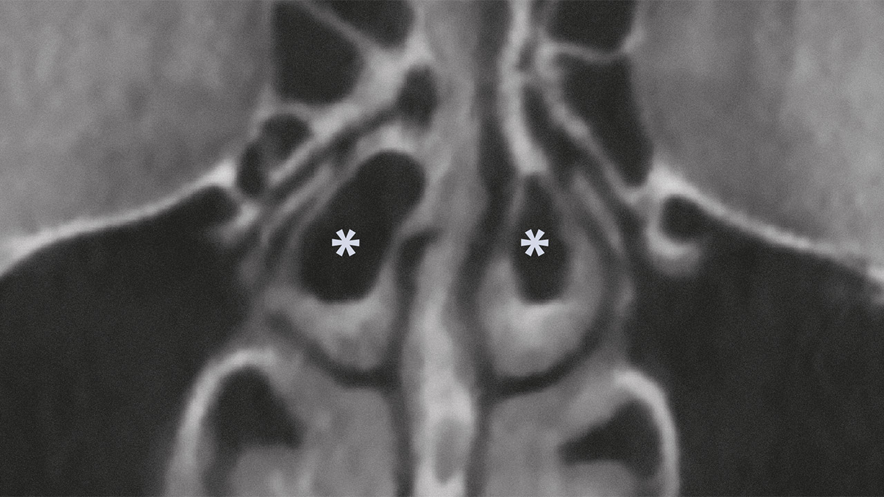
Eine FallserieMikrobielle Infektionen des Wurzelkanalsystems an Prämolaren und Molaren im Oberkiefer können im Verlauf einer periapikalen Entzündung zur Entstehung odontogener Sinusitiden führen und reaktive bzw. reparative Knochenneubildungen hervorrufen. Für eine exakte Diagnostik empfiehlt sich die Anwendung der DVT mit der vergleichenden Abbildung der rechten und linken Kieferhöhle zur Differenzialdiagnostik einer odontogenen von einer rhinogenen oder kombinierten Ursache der Sinusitis. Eine chirurgische Intervention mit Biopsie und Keimbestimmung in der Kieferhöhle sollte bei Vorliegen einer apikalen Parodontitis erst nach einem Kontrollzeitraum von 2 Jahren nach Wurzelkanalbehandlung oder bei fortbestehender Symptomatik erfolgen.
Schlagwörter: reaktive Osteogenese, Ossifikation Kieferhöhle, odontogene Sinusitis, DVT
Quintessenz Zahnmedizin, 1/2024
Bildgebende VerfahrenSeiten: 57-58, Sprache: DeutschSchulze, Dirk
AtlasQuintessenz Zahnmedizin, 9/2023
Bildgebende VerfahrenSeiten: 738-739, Sprache: DeutschSchulze, Dirk
AtlasQuintessenz Zahnmedizin, 7/2023
Bildgebende VerfahrenSeiten: 620, Sprache: DeutschSchulze, Dirk
AtlasQuintessenz Zahnmedizin, 6/2023
Bildgebende VerfahrenSeiten: 524, Sprache: DeutschSchulze, Dirk
AtlasQuintessenz Zahnmedizin, 5/2023
Bildgebende VerfahrenSeiten: 442-443, Sprache: DeutschSchulze, Dirk
Atlas