Auf unserer Website kommen verschiedene Cookies zum Einsatz: Technisch notwendige Cookies verwenden wir zu dem Zweck, Funktionen wie das Login oder einen Warenkorb zu ermöglichen. Optionale Cookies verwenden wir zu Marketing- und Optimierungszwecken, insbesondere um für Sie relevante und interessante Anzeigen bei den Plattformen von Meta (Facebook, Instagram) zu schalten. Optionale Cookies können Sie ablehnen. Mehr Informationen zur Datenerhebung und -verarbeitung finden Sie in unserer Datenschutzerklärung.
Dr. Tiziano Testori ist Leiter der Abteilung für Implantologie und orale Rehabilitation der Zahnmedizinischen Klinik am Istituto Ortopedico Galeazzi, Abteilung für Gesundheitstechnologien der Universität Mailand. Außerdem ist er Gastprofessor am New York University College of Dentistry in New York. Der ehemalige Präsident der Italienischen Gesellschaft für Oralchirurgie und Implantologie (SICOI) ist Gründungsmitglied der Advanced Implantology Study Group (AISG), Autor von mehr als 200 wissenschaftlichen Artikeln und Mitherausgeber von Sinusbodenaugmentation. Chirurgische Techniken und alternative Konzepte (Quintessenz, 2010). Dr. Testori betreibt eine Privatpraxis in Como.
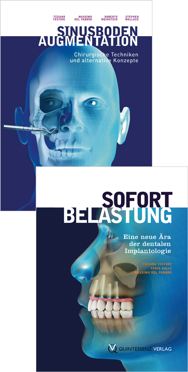
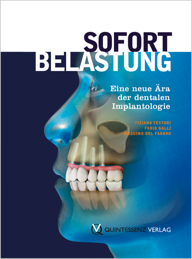
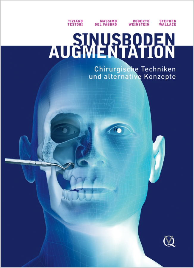
1. Auflage 2013 Buch 2 Bücher im Hardcover im Set, 976 Seiten, 1440 Abbildungen Sprache: Deutsch Kategorien: Implantologie, Prothetik, Literatur fürs Studium Artikelnr.: 17920 ISBN 978-3-86867-172-8 QP Deutschland
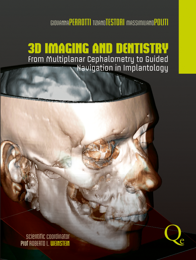
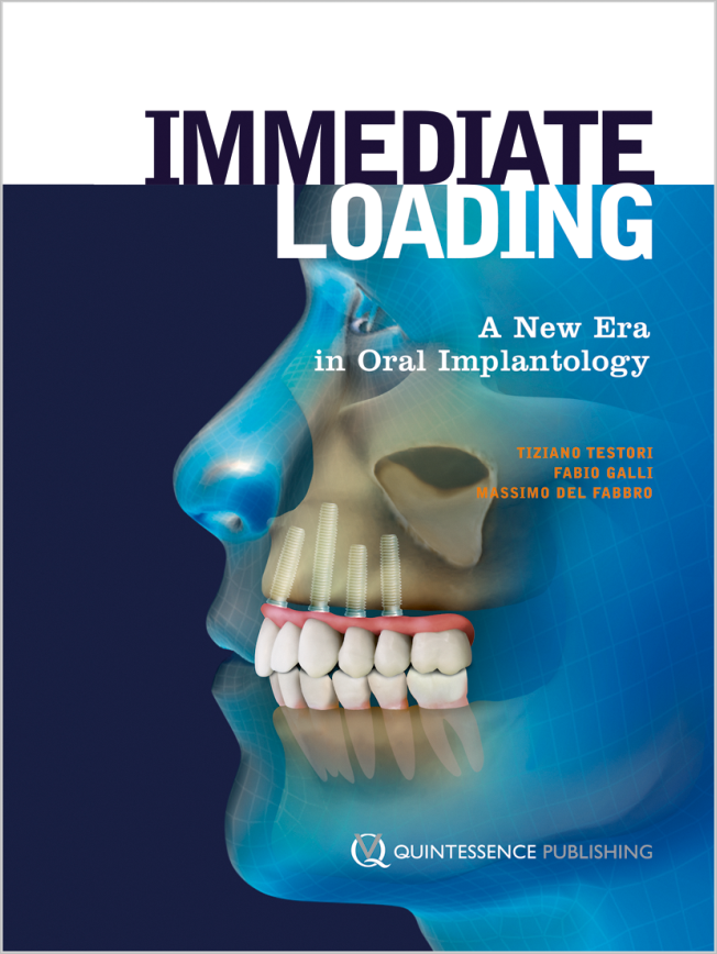
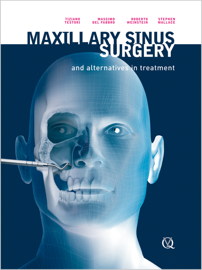
1st Edition 2009 Buch Hardcover, 21 x 28 cm, 380 Seiten, 409 farbige Abbildungen Sprache: Englisch Kategorien: Implantologie, Oralchirurgie, Parodontologie Artikelnr.: 14431 ISBN 978-1-85097-170-2 QP Deutschland
198,00 €148,00 €
Veranstaltungen
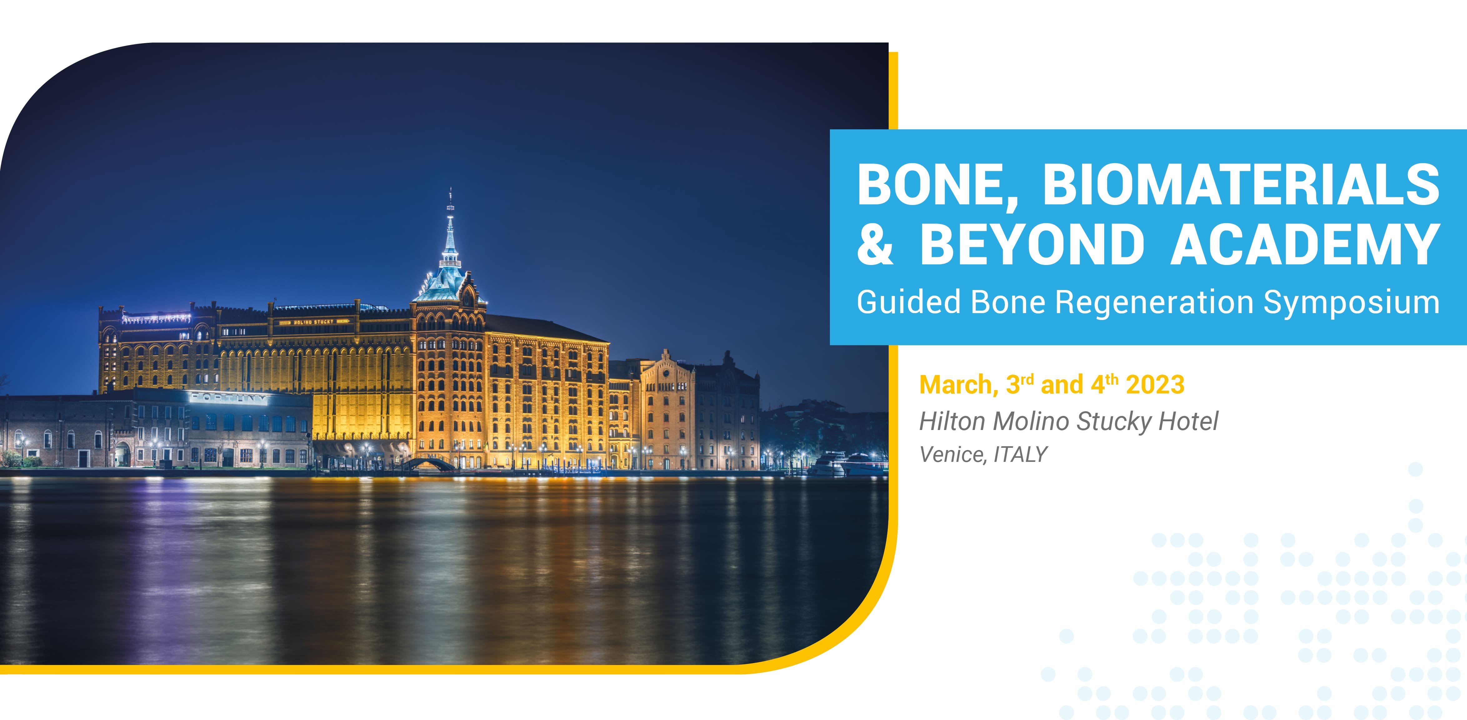
Bone, Biomaterials & Beyond Academy
Guided Bone Regeneration Symposium3. März 2023 — 4. März 2023Hilton Molino Stucky Venice, Venice, Italien
Referenten: Luca Comuzzi, Massimo del Fabbro, Abdelsalam Elaskary, Kai Fischer, Fernando Guerra, Arndt Happe, Göran Kjeller, Gerd Körner, Diego Lops, Christian Maischberger, Magda Mensi, Ulf Nannmark, Patrick Palacci, Stefano Parma-Benfenati, Adriano Piattelli, Claudio Stacchi, Tiziano Testori, Hannes Wachtel, Michael Weinländer
Bone, Biomaterials & Beyond Academy
The 14th International Symposium on Periodontics and Restorative Dentistry (ISPRD)
9. Juni 2022 — 12. Juni 2022Boston Marriott Copley Place, Boston, MA, Vereinigte Staaten von Amerika
Referenten: Tara Aghaloo, Edward P. Allen, Evanthia Anadioti, Wael Att, Vinay Bhide, Markus Blatz, Scotty Bolding, Lorenzo Breschi, Jeff Brucia, Daniel Buser, Luigi Canullo, Daniele Cardaropoli, Stephen J. Chu, Donald Clem, Christian Coachman, Lyndon F. Cooper, Daniel Cullum, Lee Culp, Sergio De Paoli, Marco Degidi, Nicholas Dello Russo, Serge Dibart, Joseph P. Fiorellini, Mauro Fradeani, Stuart J. Froum, David Garber, Maria L. Geisinger, William Giannobile, Luca Gobbato, Ueli Grunder, Galip Gürel, Chad Gwaltney, Christoph Hämmerle, Robert A. Horowitz, Markus B. Hürzeler, David Kim, Greggory Kinzer, Christopher Köttgen, Ina Köttgen, Purnima S. Kumar, Burton Langer, Lydia Legg, Pascal Magne, Kenneth A. Malament, Jay Malmquist, George Mandelaris, José Carlos Martins da Rosa, Pamela K. McClain, Michael K. McGuire, Mauro Merli, Konrad H. Meyenberg, Craig Martin Misch, Julie A. Mitchell, Marc L. Nevins, Myron Nevins, Michael G. Newman, Miguel A. Ortiz, Jacinthe M. Paquette, Stefano Parma-Benfenati, Michael A. Pikos, Giulio Rasperini, Pamela S. Ray, Christopher R. Richardson, Isabella Rocchietta, Marisa Roncati, Marco Ronda, Paul S. Rosen, Maria Emanuel Ryan, Irena Sailer, Maurice Salama, David M. Sarver, Takeshi Sasaki, Todd Scheyer, Massimo Simion, Michael Sonick, Sergio Spinato, Dennis P. Tarnow, Lorenzo Tavelli, Douglas A. Terry, Tiziano Testori, Carlo Tinti, Istvan Urban, Hom-Lay Wang, Robert Winter, Giovanni Zucchelli
Quintessence Publishing Co., Inc. USA
Zeitschriftenbeiträge dieses Autors
Quintessence International, 4/2023
DOI: 10.3290/j.qi.b3819543, PubMed-ID: 36651073Seiten: 302-318, Sprache: EnglischFischer, Kai R / Scaini, Riccardo / Chackartchi, Tali / Solderer, Alex / Schmidlin, Patrick R / Testori, Tiziano
Implant-based rehabilitation is a clinical challenge, especially in the esthetic area. Numerous factors influence the outcome of the rehabilitation; however, the two main factors are the bone and soft-tissue deficiencies at the intended implant site. Peri-implant soft tissue complications can arise from a combination of factors that can be summarized as two categories: diagnostic errors and surgical planning management errors. Most of the complications can be corrected after each step of the treatment and even after the delivery of the prosthetic restoration with adequate soft tissue management in order to give the patient an esthetically pleasing outcome. The aims of this article were: to present the current literature, to propose a clinical checklist to guide clinicians in evaluating the prognosis of the treatment utilizing soft tissue grafting, and to illustrate a case series partially employing the proposed clinical checklist. The proposed checklist could be helpful in evaluating the prognosis of the treatment utilizing only soft tissue grafting. In clinical cases in which the prognosis is classified as good, soft tissue management could be a viable treatment option before attempting more radical procedures like implant removal.
Schlagwörter: connective tissue graft, coronal advanced flap, dental implant, mucosal recession, peri-implant soft tissue deficiency, peri-implant soft tissue recession, plastic peri-implant surgery, tunnel technique
Recently, the Low Window technique was proposed to facilitate sinus augmentation and reduce postsurgical patient discomfort. It was shown to be both safe and effective. This case series evaluates the postsurgical discomfort of patients undergoing Low Window sinus augmentation, the bone gain achieved, and whether these factors correlate with the osteotomy area. Records of patients (n = 26) who underwent a sinus elevation using the Low Window approach (n = 34 interventions), followed by implant placement and prosthetic rehabilitation (n = 97 implants), were assessed retrospectively. The analyzed outcomes were the medial, middle, and lateral bone gain ≥ 9 months after augmentation; pain at 5 to 6 hours postsurgery, evaluated on a visual analog scale (VAS; 0 to 100); pain, swelling, and hematoma formation every day up to 7 days postsurgery, evaluated by VAS; implant and prosthetic success and survival rates; and rate of complications. The average follow-up time was 62.1 ± 14.9 months. No intraoperative sinus membrane perforations or other complications occurred. Medial, middle, and lateral bone gains were 10.1 ± 1.7 mm, 11.6 ± 0.8 mm, and 10.7 ± 0.9 mm, respectively. At the last follow-up, the implant success rate was 99%. Postsurgical VAS pain 5 to 6 hours postsurgery was 12.3 ± 8.0 and decreased significantly thereafter. Swelling prevalence was 29.4% at 1 day postsurgery, 20.6% at 2 days, and 2.9% the following day. No swelling was observed from day 4 onward. Prevalence of hematoma was 8.8% for 2 days postsurgery, 2.9% the day after, and no hematoma was observed thereafter. Only the osteotomy area correlated with immediate postsurgical pain, but not with other discomfort outcomes or with bone gain. Low Window sinus elevation might allow bone gain and medium-term implant and prosthetic success rates similar to that of other access window designs. Prospective, comparative studies are needed to investigate whether the technique is more advantageous than traditional approaches.
Common challenges encountered for atrophic maxilla rehabilitation are the inadequate width and height of attached keratinized mucosa (AKM) and shallow vestibular depth. This study presents a buccally displaced palatal (BDP) flap technique to increase the tissue thickness and AKM width at the second-stage surgery and reestablish the correct fornix depth. The peri-implant pocket depths, modified Plaque Index score, modified sulcus Bleeding Index score, and soft tissue recession were evaluated 6 and 12 months after prostheses loading. A total of 52 implants were placed and analyzed, and no implant failures were found. No significant changes in peri-implant parameters were observed between 6 and 12 months, and mean recession was less than 0.2 mm after 12 months. Though this change was statistically significant, it was clinically irrelevant. The results demonstrate that adequately healthy peri-implant soft tissues and substantial dimensional stability of vestibular soft tissues at the 1-year follow-up were achieved with the BDP flap technique. The BDP flap could represent a viable option for increasing the width and the height of AKM and establishing the correct maxillary fornix depth.
Maxillary sinus wall fenestration at the lateral wall or floor of the sinus can result from many potential factors, such as the repair of oro-antral communication, Caldwell-Luc antrostomy, tooth extraction after an endodontic or periodontal infection that eroded the sinus wall, and the combination of sinus pneumatization and alveolar ridge resorption after teeth removal. When sinus wall fenestration is observed on radiographs, it usually indicates adhesion between the sinus membrane and buccal flap, which makes the reentry surgery for subsequent sinus augmentation challenging. To minimize surgical complications in these challenging scenarios, this paper presents a split-flap surgical technique for the management of soft tissue adhesion between the sinus membrane and alveolar mucosa when attempting a lateral window sinus augmentation.
The aim of the present study was to generate an international and multidisciplinary consensus on the clinical management of implant protrusion into the maxillary sinuses and nasal fossae. A total of 31 experts participated, 23 of whom were experts in implantology (periodontologists, maxillofacial surgeons and implantologists), 6 were otolaryngologists and 2 were radiologists. All the participants were informed of the current scientific knowledge on the topic based on a systematic search of the literature. A list of statements was created and divided into three surveys: one for all participants, one for implant providers and radiologists and one for otolaryngologists and radiologists. A consensus was reached on 15 out of 17 statements. According to the participants, osseointegrated implants protruding radiographically into the maxillary sinus or nasal fossae require as much monitoring and maintenance as implants fully covered by bone. In the event of symptoms of sinusitis, collaboration between implant providers and otolaryngologists is required. Implant removal should be considered only after pharmacological and surgical management of sinusitis have failed.
Schlagwörter: consensus, dental implants, maxillary sinus, nasal fossae
Conflict-of-interest statement: The authors declare there are no conflicts of interest relating to this study.
Purpose: To assess whether bone grafting is associated with early implant failure (defined as a lack of osseointegration detected prior to functional loading) and to evaluate the association between bone grafting procedures and other risk factors for early implant failure.
Materials and methods: Two independent reviewers conducted an electronic search of MEDLINE (via PubMed). Meta-analysis was performed for the odds ratio of bone grafting procedures associated with early implant failure. The Newcastle-Ottawa Scale for cohort studies was used to assess the risk of bias.
Results: Of the 231 articles selected for full-text review, 10 were included in the qualitative analysis and for quantitative meta-analysis. An odds ratio of 1.50 (95% confidence interval 1.06–2.13) was recorded for bone grafting procedures associated with early implant failure. Data regarding the association of bone grafting and other risk factors in determining early implant failure were insufficient for quantitative analysis.
Conclusions: Within the limitations of this study, a significant positive association was found between bone grafting procedures and early implant failure. The possible negative effect of bone grafting procedures on implant osseointegration should be considered when planning implant therapy.
Schlagwörter: alveolar bone grafting, dental implants, early implant failure, meta-analysis, systematic review
Conflict-of-interest statement: The authors declare there are no conflicts of interest relating to this study.
Purpose: The aim of this clinical study was to verify the predictability of the rehabilitation of extremely atrophic jaws with immediately loaded short implants and evaluate posterior mandibular regrowth.
Materials and methods: A cohort of consecutive fully edentulous patients wearing complete dentures in both arches was enrolled. Periodically, implant survival and prosthetic success were assessed. After informed consent, a subsample of 10 patients who had preoperative CBCT underwent a postoperative CBCT 1 year after immediate implant loading, and 3D superimpositions of pre- and postoperative images were performed. Linear measurements of bone height were performed at two sites in each hemimandible and, on the same sections, bone density according to the qualitative gray values (GVs) was analyzed in an area of 3 mm2 including the cortical mandibular bone.
Results: Fifty-nine fully edentulous patients (31 females, 28 males) with Class VI atrophic mandibles according to Cawood and Howell were rehabilitated with the insertion of four to five short implants (4-mm diameter, 7- or 8.5-mm long). Overall, 251 implants were immediately loaded with a fixed hybrid prosthesis. Four patients did not show up for recall visits, bringing the final number down to 55 patients (31 females, 24 males) and 231 implants. In up to 14 years of follow-up, a total of 4 implant failures were recorded (cumulative survival rate, 98.4%). The biologic complications included 9 mucositis (3.9%) and 3 peri-implantitis (1.3%) at implant-level analysis. Mechanical complications involved 9 chippings of the prosthetic restorations (17.0%). In the 10-patient subsample, the analysis showed bone growth (average of 1.2 ± 0.7 mm) in the posterior areas of the mandible. In addition, bone density was found to increase 17% to 27% with reference to the preoperative CBCT.
Conclusion: The immediate loading of short implants may represent a feasible therapeutic option for the treatment of fully edentulous patients with severely atrophic mandibles. Bone regrowth in the posterior areas and an increase in bone density of the mandible may occur.
Schlagwörter: atrophic mandibles, immediate implant loading, implant survival
Maxillary sinus grafting is generally a safe procedure. However, intraoperative complications, as well as early and late postoperative complications, may occur. Included in the latter group are graft infections that can be triggered by peri-implantitis. The aim of the present study was to report three cases of late maxillary sinus graft infections and to histologically evaluate the effects of peri-implantitis in the grafted area. In peri-implantitis cases in grafted sinuses, the sole removal of the implant along with accompanying debridement of the infected area may not be sufficient to resolve the infection, and a more-aggressive treatment may be necessary.
This study aimed to evaluate the correlation between soft tissue thickness measured by CBCT and phenotype probing estimation and to assess the thickness cutoffs for each phenotype probing outcome. CBCT was performed with a lip retractor in order to isolate periodontal soft tissues in 10 consecutive patients. Using colored probes, the phenotype was evaluated for all present teeth and recorded as thin, medium, thick, or very thick. The overall correlation between tissue thickness and the phenotype probe score was r = 0.86 (CI: 0.80, 0.90). The correlation was r = 0.90 (CI: 0.81, 0.94) when only maxillary anterior teeth were considered. The obtained cutoffs were 0.83 mm between thin and medium phenotypes, 1.07 mm between medium and thick phenotypes, and 1.24 mm between thick and very thick phenotypes. Thus, a high correlation between tissue thickness and the phenotype probe score was found. Preliminary data on the use of phenotype probes as an evaluation method for gingival thickness were promising.
The aim of this article is to propose a simplified digital protocol for the treatment of the fully edentulous patient, using an immediate implant and immediate loading protocol to deliver a polymethyl methacrylate metal-reinforced hybrid prosthesis. Ten consecutive patients were treated with this approach. At the end of 1 year, there was an implant survival rate of 97.8% and a prosthetic success rate of 100%. Based on the responses to the quality of life questionnaire, patients had a high acceptance rate for this treatment protocol. Within the limits of this case series, the proposed simplified digital protocol could be utilized for reconstruction in the fully edentulous patient and for delivering an implant-supported prosthesis immediately after implant placement.