International Journal of Periodontics & Restorative Dentistry, Pre-Print
DOI: 10.11607/prd.6626, PubMed-ID: 3781984911. Okt. 2023,Sprache: EnglischFonseca, Manrique / Molinero-Mourelle, Pedro / Donmez, Mustafa Borga / Abou-Ayash, Samir / Buser, Daniel / Sculean, Anton / Yilmaz, Burak
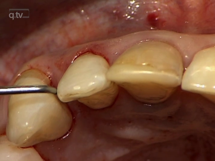
Dental implants have been commonly used to replace missing single teeth. However, esthetic rehabilitation of an adjacent tooth may also be required due to diastemas, crowding, or existing large direct restorations to improve the final esthetic outcome. With the advancements in ceramics and bonding techniques, minimally invasive esthetic approaches have become viable for compromised spacing issues. This case report describes a dental technique for the esthetic rehabilitation of compromised anterior spacing with a customized zirconia implant abutment at maxillary central incisor site and a partial ceramic veneer bonded to adjacent central incisor.
Schlagwörter: Anterior spacing; case report; implants; partial laminate veneer; prosthetic dentistry
Oral Health and Preventive Dentistry, 1/2024
Open Access Online OnlyOral HealthDOI: 10.3290/j.ohpd.b4997035, PubMed-ID: 38376435Seiten: 115-122, Sprache: EnglischWolf, Thomas Gerhard / Dianišková, Simona / Cavallé, Edoardo / Aliyeva, Rena / Cagetti, Maria-Grazia / Campus, Guglielmo / Deschner, James / Forna, Norina / Ilhan, Duygu / Mazevet, Marco / Lella, Anna / Melo, Paulo / Perlea, Paula / Rovera, Angela / Sculean, Anton / Sharkov, Nikolai / Slutsky, Ariel / Torres, António Roma / Saag, Mare
Purpose: Dental students learn knowledge and practical skills to provide oral health care to the population. Practical skills must be maintained or continuously developed throughout a professional career. This cross-sectional survey aimed to evaluate the perception of practical skills of dental students and dental-school graduates by national dental associations (NDAs) in international comparison in the European Regional Organization of the FDI World Dental Federation (ERO-FDI) zone.
Materials and Methods: A questionnaire of 14 items collected information on pre-/postgraduate areas.
Results: A total of 25 countries participated (response rate: 69.4%), with 80.0% having minimum requirements for practical skills acquisition and 64.0% starting practical training in the 3rd year of study. In countries where clinical practical work on patients begins in the 2nd year of study, practical skills of graduates are perceived as average, starting in the 3rd year of study as mainly good, starting in the 4th as varying widely from poor to very good. In total, 76.0% of respondents feel that improvements are needed before entering dental practice. Improvements could be reached by treating more patients in dental school (32.0%), increasing the quantity of clinical training (20.0%), or having more clinical instructors (12.0%). In 56.0% of the countries, it is possible to open one’s own dental practice immediately after graduation, and in 16.0%, prior vocational training is mandatory.
Conclusions: All participating countries in the ERO-FDI zone reported practical training in dental school, most starting in the 3rd year of study. The perception of practical skills of dental students and dental-school graduates among NDAs is very heterogeneous. Reasons for the perceived deficiencies should be further explored.
Schlagwörter: dental association, graduate, international, practical skills, student
Quintessence International, 9/2023
DOI: 10.3290/j.qi.b4171703, PubMed-ID: 37345441Seiten: 712-722, Sprache: EnglischKauffmann, Frederic / Fickl, Stefan / Sculean, Anton / Fischer, Kai R. / Friedmann, Anton
Objective: To clinically and histologically evaluate the potential effect of a cross-linked, high molecular weight hyaluronic acid (xHyA) on the outcomes of guided bone regeneration performed with a demineralized bovine bone mineral (DBBM) covered with a natural collagen membrane.
Method and materials: Eleven patients (eight females and three males, mean age 53 years) with a total of 27 surgical sites were treated. Treatments were performed with either DBBM and natural collagen membrane fixed with tacks (group A) or DBBM mixed with xHyA and subsequently covered with natural collagen membrane (group B). Clinical evaluations were made at baseline (T1), immediately after guided bone regeneration (T2), and at the time of implant placement (T3). Additionally, at the time of implant placement, core biopsies were retrieved and submitted for histologic analysis.
Results: Healing was uneventful in all cases. At 6 months, group B revealed a statistically significantly higher crestal ridge dimension compared to group A (P = .007). The histologic analysis revealed a tendency for greater mineralized tissue formation in group B compared to group A (67.5% versus 41.6%) and contained a higher amount of new bone (37.2%) and less DBBM residues (20.9%) than group A (12.8% new bone and 28.8% DBBM residues, respectively).
Conclusions: Within their limits, the present data indicate that, during guided bone regeneration with natural collagen membrane, the combination of DBBM and xHyA may improve the quality and quantity of bone formed with DBBM alone.
Schlagwörter: bone defect, bone grafting, bovine bone–derived mineral, cross-linked hyaluronic acid, graft fixation, graft stability, guided bone regeneration, histomorphometry, resorbable membrane
Quintessence International, 8/2023
DOI: 10.3290/j.qi.b4007601, PubMed-ID: 37010441Seiten: 622-628, Sprache: EnglischChackartchi, Tali / Imber, Jean-Claude / Stähli, Alexandra / Bosshardt, Dieter / Sacks, Hagit / Nagy, Katalin / Sculean, Anton
Objective: To histologically evaluate the effects of a novel human recombinant amelogenin (rAmelX) on periodontal wound healing/regeneration in intrabony defects.
Method and materials: Intrabony defects were surgically created in the mandible of three minipigs. Twelve defects were randomly treated with either rAmelX and carrier (test group) or with the carrier only (control group). At 3 months following reconstructive surgery, the animals were euthanized, and the tissues histologically processed. Thereafter, descriptive histology, histometry, and statistical analyses were performed.
Results: Postoperative clinical healing was uneventful. At the defect level, no adverse reactions (eg, suppuration, abscess formation, unusual inflammatory reaction) were observed with a good biocompatibility of the tested products. The test group yielded higher values for new cementum formation (4.81 ± 1.17 mm) compared to the control group (4.39 ± 1.71 mm) without reaching statistical significance (P = .937). Moreover, regrowth of new bone was greater in the test compared to the control group (3.51 mm and 2.97 mm, respectively, P = .309).
Conclusions: The present results provided for the first-time histologic evidence for periodontal regeneration following the use of rAmelX in intrabony defects, thus pointing to the potential of this novel recombinant amelogenin as a possible alternative to regenerative materials from animal origins.
Schlagwörter: amelogenin, enamel matrix derivative, intrabony defects, periodontal regeneration, recombinant, wound healing
Quintessence International, 5/2023
DOI: 10.3290/j.qi.b3824933, PubMed-ID: 36661359Seiten: 384-392, Sprache: EnglischRoccuzzo, Andrea / Ettmayer, Johanna / De Ry, Siro Pietro / Imber, Jean-Claude / Sculean, Anton / Salvi, Giovanni Edoardo
Objectives: To assess the association between the baseline radiographic defect angle and the long-term clinical outcomes following periodontal regenerative therapy with enamel matrix derivative (EMD).
Method and materials: Baseline periapical radiographs obtained from a cohort of patients treated with periodontal regenerative therapy were digitized and the radiographic angle width between the root surface and the bony wall of the adjacent intraosseous defect was calculated and reported (in degrees). Changes in pocket probing depth (PD) and clinical attachment level (CAL) were assessed and reported (in mm). Clinical outcomes were evaluated at baseline (T0), 6 months following therapy (T1), and at the latest follow-up (T2).
Results: Thirty-eight defects in 26 patients enrolled in supportive periodontal care for a mean period of 10.4 years (range 8.0 to 15.5 years) were available for analysis. The mean PD change between T0 and T2 was 2.33 ± 1.66 mm at teeth with a defect angle width < 20 degrees and 0.86 ± 1.66 mm at teeth with a defect angle width > 30 degrees (P = .021). When the baseline radiographic angle width was < 20 degrees the probability of obtaining a CAL gain > 3 mm was 1.5-times higher (95% CI 0.19 to 13.8) at T1 and 2.5-times higher (95% CI 0.40 to 15.6) at T2 compared with defects with a radiographic angle width > 30 degrees.
Conclusion: Within their limitations, these results indicate that pretherapeutic measurement of the radiographic defect angle width might provide relevant information on the short-/long-term clinical outcomes following regenerative periodontal therapy with EMD.
Schlagwörter: enamel matrix derivative, intrabony defects, long-term results, periodontal regeneration, radiographic evaluation
International Journal of Oral Implantology, 3/2023
PubMed-ID: 37767616Seiten: 211-222, Sprache: EnglischRoccuzzo, Andrea / Weigel, Lucienne / Marruganti, Crystal / Imber, Jean-Claude / Ramieri, Guglielmo / Sculean, Anton / Salvi, Giovanni E / Roccuzzo, Mario
Purpose: To longitudinally assess the prevalence of peri-implant health, peri-implant mucositis and peri-implantitis in a cohort of patients with and without history of periodontitis over a 20-year period.
Materials and methods: Eighty-four patients who attended a specialist private periodontal practice were evaluated prospectively 10 and 20 years after prosthesis delivery. Following successful completion of periodontal/implant therapy, patients (172 implants) were enrolled on an individualised supportive periodontal care programme. Clinical and radiographic parameters were collected to assess the prevalence of peri-implant health and diseases. Prevalence of peri-implantitis and peri-implant mucositis was calculated based on the case definition set out in 2018. A multilevel logistic regression analysis was conducted to assess potential risk or protective factors.
Results: The analysis was performed on 22 periodontally healthy and 62 periodontally compromised patients rehabilitated with 39 and 130 implants, respectively. The 10-year prevalence of peri-implant health, peri-implant mucositis and peri-implantitis was 21.4%, 67.9% and 10.6%, respectively, whereas the 20-year prevalence was 29.8%, 47.6% and 33.3%, respectively. Non-compliant periodontally compromised patients showed a statistically significantly increased risk at 20 years of both peri-implant mucositis (odds ratio 11.1; 95% confidence interval 1.8–68.6) and peri-implantitis (bone loss and probing depth) (odds ratio 14.3; 95% confidence interval 1.8–32.9). High full-mouth plaque and bleeding scores were associated with higher odds of both peri-implant mucositis and peri-implantitis.
Conclusions: Peri-implant diseases were prevalent in patients rehabilitated with dental implants and followed up for a period of 20 years. History of periodontal disease and a lack of compliance with a tailored supportive periodontal care programme were identified as risk factors for peri-implant diseases.
Schlagwörter: dental implants, peri-implantitis, periodontally compromised patients, periodontitis, supportive periodontal care
The authors declare no potential conflict of interests with respect to this study. The study was self-funded; no external funding was available for this research.
Oral Health and Preventive Dentistry, 1/2023
Open Access Online OnlyPeriodontologyDOI: 10.3290/j.ohpd.b4347453, PubMed-ID: 3772489730. Aug. 2023,Seiten: 279-284, Sprache: EnglischRamanauskaite, Egle / Machiulskiene, Vita / Dvyliene, Urte Marija / Eliezer, Meizi / Sculean, Anton
Purpose: The adjunctive subgingival application of sodium hypochlorite/amino acid and a mixture of natural and cross-linked hyaluronic acid gels (high molecular weight) has been recently proposed as a novel modality to enhance the outcomes of non-surgical periodontal therapy. The aim of this prospective case series was to evaluate the clinical outcomes obtained following the subgingival application of a combination of sodium hypochlorite/amino acid and a mixture of natural and cross-linked hyaluronic acid (high molecular) gels in conjunction with non-surgical periodontal therapy. Material and Methods: Twenty-one systemically healthy, non-smoking patients diagnosed with stage II-III, grade A/B periodontitis underwent full-mouth subgingival debridement (SD) performed with ultrasonic and hand instruments. All sites with probing depths (PD) ≥ 4 mm were treated with additional repeated (i.e., 2-3 times) instillation of sodium hypochlorite/amino acid gel in the periodontal pockets prior to and during SRP. Following mechanical debridement, a mixture of natural and cross-linked hyaluronic acid (high molecular) gel was applied in the pockets. The primary outcome variable was PD reduction; changes in clinical attachment level (CAL) and bleeding on probing (BOP) were the secondary outcomes. The clinical parameters were assessed at baseline, 3 and 6 months after therapy.
Results: Compared to baseline, a statistically significant mean reduction of PD values was obtained after 3 and 6 months, amounting to 2.6 ± 0.4 mm, and 2.9 ± 0.4 mm, respectively (p < 0.001). Mean CAL gain measured 2.3 ± 0.5 mm at 3 months and 2.6 ± 0.5 mm at 6 months in comparison to baseline (p < 0.001). Mean reduction of BOP values was 54.9 ± 16.9 % at 3 months and 65.6 ± 16.4 % at 6 months (p < 0.001). The number of moderate pockets (4-5 mm) decreased from 1808 at baseline to 274 at the 6-month evaluation, and the number of deep (≥ 6 mm) pockets dropped from 319 to 3, respectively.
Conclusion: The combination of sodium hypochlorite/amino acid and a mixture of natural and cross-linked hyaluronic acid (high molecular) adjunctive to subgingival debridement may represent a valuable approach to improve the outcomes of non-surgical periodontal treatment.
Schlagwörter: cross-linked hyaluronic acid, non-surgical periodontal therapy, periodontitis, sodium hypochlorite/amino acid.
Oral Health and Preventive Dentistry, 1/2023
Open Access Online OnlyPeriodontologyDOI: 10.3290/j.ohpd.b4009557, PubMed-ID: 370142144. Apr. 2023,Seiten: 103-112, Sprache: EnglischKardaras, Giorgios / Marcovici, Iasmina / Rusu, Darian / Dehelean, Cristina / Coricovac, Dorina / Iorio-Siciliano, Vincenzo / Sculean, Anton / Stratul, Stefan-Ioan
Purpose: Since NaOCl acts as a strong oxidizing agent and presents potential toxicity, this study was adressed to evaluate the in-vitro safety of NaOCl solutions at concentrations below the limit of patient tolerance, i.e. ≥ 0.5%.
Materials and Methods: First, an in-silico evaluation was conducted to predict the potential toxicity of NaOCl in terms of mutagenic, tumorigenic, irritant, and reproductive risks, as well as some drug-like properties of the molecule. The in-vitro experiments were based on 2D and 3D models. For the 2D approach, two selected cell lines – HaCaT (human skin keratinocytes) and HGF (human gingival fibroblasts) – were exposed to NaOCl at five concentrations (0.05 – 0.5%) for 10, 30, and 60 s to simulate possible clinical administration. The irritative potential of NaOCl 0.05% and 0.25% was assessed in a 3D in-vitro model (EpiDerm, reconstructed human epidermis). Statistical significance was set at p < 0.05.
Results: The main findings suggest that NaOCl exerts cytotoxicity towards HaCaT immortalised keratinocytes and HGF primary gingival fibroblasts in a cell type-, dose- and time-dependent manner, with the most prominent effect being recorded in HaCaT cells after 60 s of treatment with NaOCl 0.5%. However, NaOCl was computationally predicted as free of mutagenic, tumorigenic, irritant, and reproductive toxicity, and showed no irritative potential in 3D reconstructed epidermis at concentrations of 0.05% and 0.25%.
Conclusion: Further clinical and histological studies are required to confirm these results, as well as elucidate the potential cytotoxic mechanism induced by NaOCl in HaCaT and HGF cells at the tested concentrations.
Schlagwörter: periodontitis, primary gingival fibroblasts, sodium hypochlorite, toxicity
Oral Health and Preventive Dentistry, 1/2023
Open Access Online OnlyOral MedicineDOI: 10.3290/j.ohpd.b3920023, PubMed-ID: 3682564024. Feb. 2023,Seiten: 69-76, Sprache: EnglischTennert, Christian / Sarra, Giada / Stähli, Alexandra / Sculean, Anton / Eick, Sigrun
Purpose: To evaluate the effect of bovine milk and yogurt on selected oral microorganisms and different oral biofilms.
Materials and Methods: Milk was prepared from 0.5% fat (low-fat) and 16% fat (high fat) milk powder. For yogurt preparation, the strains Lactobacillus delbrueckii ssp. bulgarcius and Streptococcus thermophilus were added to the milk. Minimal inhibitory concentrations (MIC) and minimal microbiocidal concentrations (MMC) of the test compounds were measured against various microorganisms by the microbroth dilution technique. Cariogenic periodontal biofilms and one containing Candida were created on plastic surfaces coated with test substances. Further, preformed biofilms were exposed to the test substances at a concentration of 100% for 10 min and thereafter 10% for 50 min. Both colony forming units (cfu) and metabolic activity were quantified in the biofilms.
Results: Neither high-fat milk, low-fat milk nor casein inhibited the growth of any species. Yogurt and L. delbrueckii ssp. bulgaricus at low MIC and MMC suppressed the growth of Porphyromonas gingivalis and other bacteria associated with periodontal disease. High-fat yogurt decreased cfu in the forming periodontal biofilm by 90%. Both low- and high-fat yogurts reduced metabolic activity in newly forming and preformed periodontal and Candida biofilms, but not in the cariogenic biofilm.
Conclusions: Yogurt and L. delbru eckii ssp. bulgaricus, but not milk, were bactericidal against periodontopathogenic bacteria. Yoghurt reduced the metabolic activity of a Candida biofilm and a periodontal biofilm. Yogurt and L. delbrueckii ssp. bulgaricus may have potential in prevention and therapy of periodontal diseases and Candida infections.
Schlagwörter: Candida, caries, milk products, oral bacteria, periodontitis
Oral Health and Preventive Dentistry, 1/2023
Open Access Online OnlyPubMed-ID: 3734558122. Juni 2023,Seiten: 1, Sprache: EnglischSculean, Anton / Banerjee, Avijit / Rothenbücher, Marina


